
Rapid Review·Cardiovascular
Select any text to highlight it or add a note.
PATHOLOGY
T1Must knowCongenital Heart Disease: Right-To-Left Shunts
P300
Videos
5 to play here
Play 5 here
Focus on
The 5 T’s: the blue babies who need a shunt kept open just to survive.
Key takeaways
The blue babies
Right-to-left shunts cause EARLY cyanosis, the "blue babies"
usually diagnosed prenatally or apparent immediately after birth, and they typically need urgent surgery and a PDA held open with prostaglandin.The 5 T's, numbered so the mnemonic does the work
- 1. Truncus arteriosus = 1 vessel
- 2. Transposition = 2 switched vessels
- 3. Tricuspid atresia = 3, as in Tri
- 4. Tetralogy of Fallot = 4, as in Tetra
- 5. TAPVR = 5 letters
| Association | Defects |
|---|---|
| Fetal alcohol syndrome | VSD, PDA, ASD, tetralogy of Fallot |
| Congenital rubella | PDA, pulmonary artery stenosis, septal defects |
| Down syndrome | AV septal defect (endocardial cushion defect), VSD, ASD |
| Infant of a diabetic mother | Transposition of the great arteries, truncus arteriosus, tricuspid atresia, VSD |
| Marfan syndrome | MVP, thoracic aortic aneurysm and dissection, aortic regurgitation |
| Prenatal lithium exposure | Ebstein anomaly |
| Turner syndrome | Bicuspid aortic valve, coarctation of the aorta |
| Williams syndrome | Supravalvular aortic stenosis |
| 22q11 syndromes | Truncus arteriosus, tetralogy of Fallot |
The 5 T's, one by one
Persistent truncus arteriosus
the truncus never divides, because the aorticopulmonary septum fails to form, so one trunk receives the output of both ventricles. The septum fails because neural crest cells fail to migrate into the outflow tract. Most cases carry an accompanying VSD.D-transposition of the great arteries
compare the transposition diagram with the transposition on MRI.- The aorta leaves the RV (anteriorly) and the pulmonary trunk leaves the LV (posteriorly), so the systemic and pulmonary circulations run in parallel rather than in series.
- Not compatible with life unless a shunt exists, i.e. a VSD, PDA, or patent foramen ovale.
- Caused by failure of the aorticopulmonary septum to spiral.
- "Egg on a string" on chest x-ray, from the narrow superior mediastinum. Without surgery most infants die within the first few months.
- Hyperoxia test: arterial oxygenation barely rises on 100% oxygen, because the circuits do not mix. Typically no murmur, since the shunt flow is low velocity.
Tricuspid atresia
absent tricuspid valve with a hypoplastic RV and a dilated RA. Needs both an ASD and a VSD or PDA to be viable. ECG shows RA hypertrophy (tall P waves) and LV hypertrophy (left axis deviation), because the RV never developed.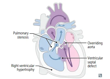
What this shows
Tetralogy of Fallot
by "PROVe", all four drawn on the figure.- Pulmonary infundibular stenosis, the most important determinant of prognosis. It forces right-to-left flow across the VSD, which is what builds the RVH.
- Right ventricular hypertrophy, giving the boot-shaped heart on the boot-shaped heart x-ray
- Overriding aorta, straddling the VSD
- VSD
- Caused by anterosuperior displacement of the infundibular septum. The most common cause of early childhood cyanosis, and associated with 22q11 syndromes.
- Called tetralogy meaning 4 things, not teratology; Fallot is just the name of the French physician who first described it.
TAPVR
the pulmonary veins drain into the right heart (for example into the SVC or coronary sinus) instead of the LA, so an ASD (with or without a PDA) is required to shunt right to left and maintain cardiac output. Rarely asked.Ebstein anomaly
Ebstein anomaly
downward displacement of the tricuspid leaflets into the RV, "atrializing" the ventricle. I don't get why it's not called "ventriculizing" of the atria because it makes more sense, but whatever. Caused by in-utero lithium exposure, which can also cause TAPVR.What it gives
tricuspid regurgitation, accessory conduction pathways (WPW), and right-sided heart failure.On auscultation
a widely split S1 (the malformed tricuspid closes late), the murmur of tricuspid regurgitation, and an early systolic click (the "sail sound" of the malformed leaflet).| Lesion | Mnemonic number | Defining defect | Requires for survival |
|---|---|---|---|
| Truncus arteriosus | 1 vessel | Aorticopulmonary septum never forms | Accompanying VSD, usually present |
| D-transposition | 2 switched vessels | Aorticopulmonary septum fails to spiral | VSD, PDA, or PFO, or it is fatal |
| Tricuspid atresia | 3 = Tri | Absent tricuspid valve, hypoplastic RV | Both ASD and VSD/PDA |
| Tetralogy of Fallot | 4 = Tetra | Anterosuperior displacement of the infundibular septum | Not duct-dependent unless severe |
| TAPVR | 5 letters | Pulmonary veins drain to the right heart | ASD, with or without PDA |
| Finding | Lesion |
|---|---|
| "Egg on a string" | D-transposition |
| "Boot-shaped heart" | Tetralogy of Fallot (RVH) |
| "Atrialized" right ventricle | Ebstein anomaly |
| Feature | Right-to-left | Left-to-right |
|---|---|---|
| Cyanosis | EARLY, the "blue babies" | Late, once Eisenmenger develops |
| Presentation | Prenatal or immediately after birth | Acyanotic at presentation |
| Management | Urgent surgery; keep the PDA open with prostaglandin | Repair according to complications |
A newborn stays cyanotic on 100% oxygen, has no murmur, and the x-ray shows a narrow superior mediastinum. Diagnosis, and what keeps the baby alive?
D-transposition of the great arteries: the circuits run in parallel, so only a shunt (VSD, PDA, or PFO) lets blood mix; keep the PDA open with prostaglandin until surgery.
How it's tested
A school-age child who squats on the playground to relieve cyanosis has tetralogy of Fallot, and squatting works by raising systemic vascular resistance. Mehlman is specific that it is the afterload effect, not the preload effect, that improves symptoms, and that is the distractor the question is built around. Of the four components, the degree of pulmonary infundibular stenosis determines prognosis, so that is the answer when asked which feature matters most.
Go deeper
First Aid 2026 — CV/Pathology (p.300) · B&B — Right-to-left shunts · Mehlman — HY Cardio (5 T’s, squatting and afterload)
High-yield images9
Flashcards for this page
Card 1 of 5 · try-out only, nothing is saved
(...)
You just read one page of 944
The rest of Step 1, written exactly like this.
Rapid Review is the reading layer of a full Step 1 platform: a schedule fitted to your exam date, flashcards for every page, and a mentor a message away.
Every Step 1 system
Written the same way: what to know, how it’s tested, where to go deeper.
A plan built to your exam date
Tell us when you sit, and the schedule fits the whole library and your question bank to it.
Flashcards for every page
Ready-made spaced-repetition decks linked to each topic, so nothing you read gets forgotten.
