
Rapid Review·Cardiovascular
Select any text to highlight it or add a note.
Congenital Heart Disease
T2High yieldEbstein Anomaly
Focus on
A tricuspid valve displaced down into the right ventricle, "atrializing" its upper part — after maternal lithium. Severe tricuspid regurgitation, a huge right atrium, cyanosis through a PFO, and Wolff-Parkinson-White in a third. The spectrum runs from hydrops in the fetus to an adult with palpitations; repair the valve when the RV fails.
Key takeaways
What it is
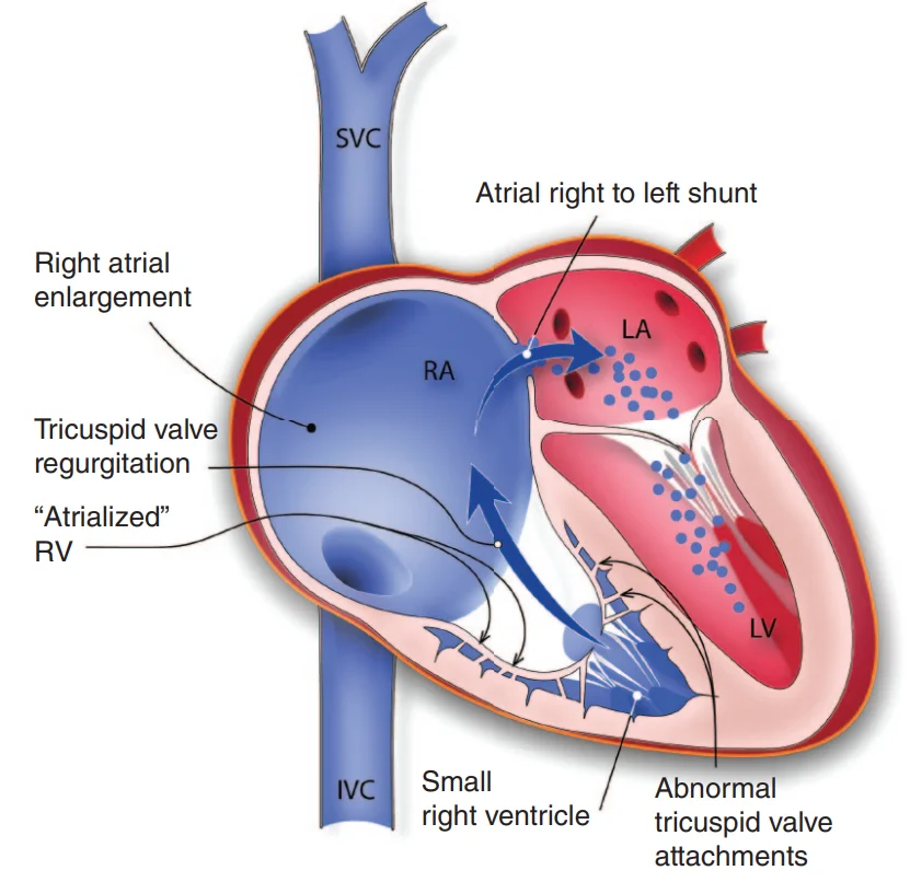
What this shows
Ebstein anomaly
a malformed, downward-displaced tricuspid valve attached to the right-ventricular wall.- The inflow portion of the ventricle becomes a thin, poorly contractile "atrialized right ventricle", the functional ventricle is small, and the valve leaks.
Consequences
tricuspid regurgitation, right-heart enlargement, right-to-left atrial shunting (cyanosis), and arrhythmias through accessory pathways (Wolff-Parkinson-White Syndrome).Association
maternal lithium use early in pregnancy.Clinical features and diagnosis
Severe disease
in-utero heart failure with non-immune hydrops fetalis and death.- Cyanosis and heart failure in the newborn, worsened by the high neonatal pulmonary resistance and often improving as it falls.
Adolescents and adults
palpitations and supraventricular tachycardia (Wolff-Parkinson-White), exertional dyspnea, cyanosis with exertion, right heart failure, paradoxical embolism.Examination
a loud S1 and a holosystolic murmur of tricuspid regurgitation at the left sternal border, widely split S1 and S2 (right-ventricular volume overload), a triple or quadruple rhythm, hepatomegaly.Transthoracic echocardiography is the best confirmatory test
apical displacement of the septal leaflet, the atrialized ventricle, tricuspid regurgitation, marked right-atrial and ventricular dilation, the atrial shunt.Electrocardiogram
tall broad P waves (right atrial enlargement), a long PR, right bundle branch block, delta waves in a third.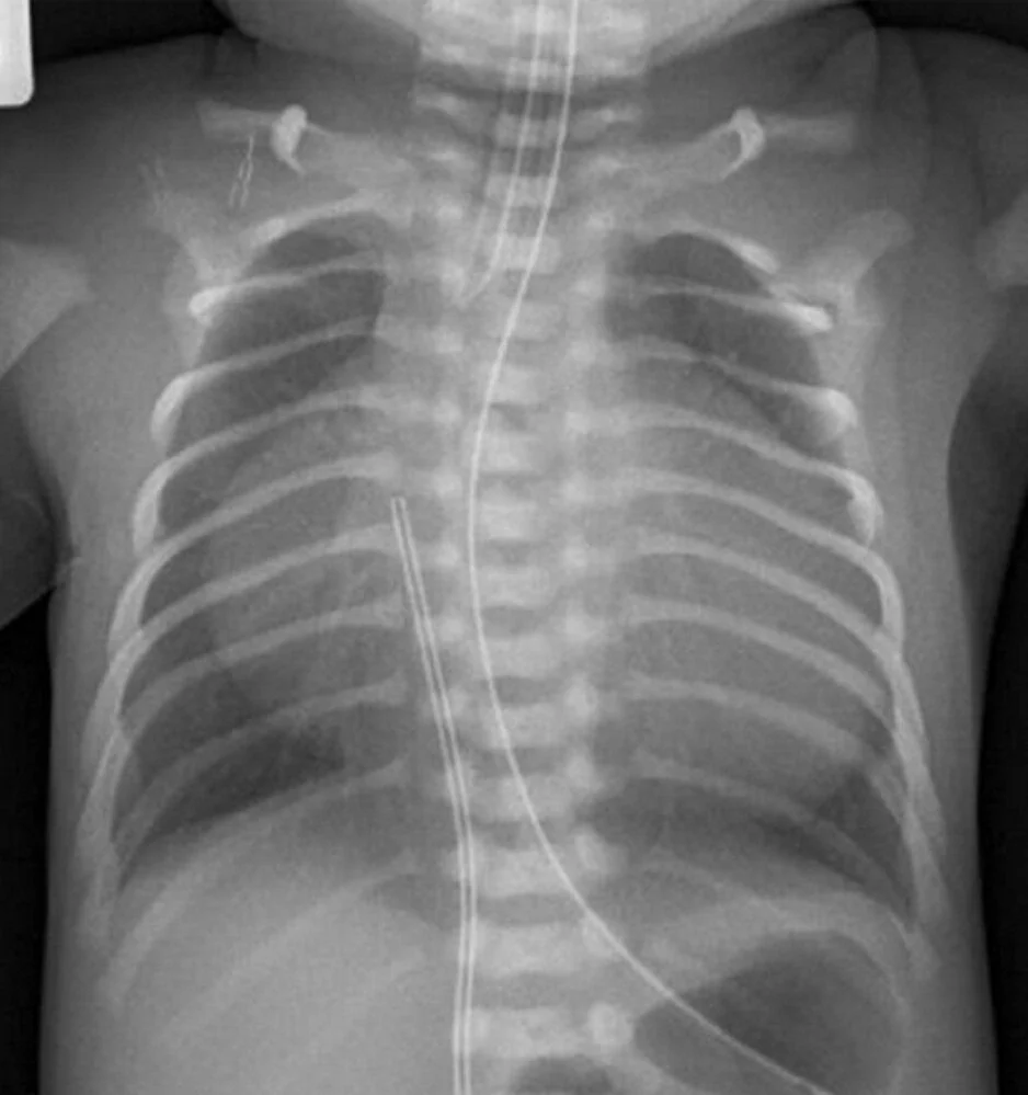
What this shows
Chest film
massive globular cardiomegaly ("wall-to-wall heart") from the huge right atrium, with decreased pulmonary vascular markings.| Ebstein anomaly | Tricuspid Atresia | Pulmonary atresia with intact septum | |
|---|---|---|---|
| Valve | Displaced, regurgitant tricuspid valve | Absent tricuspid valve | Atretic pulmonary valve |
| Right ventricle | Atrialized, small functional ventricle | Hypoplastic | Hypoplastic, hypertensive |
| Heart size on film | Massive cardiomegaly | Small to normal | Variable |
| Electrocardiogram | Tall P, right bundle branch block, long PR, pre-excitation | Left axis deviation | Right axis |
| Key clue | Maternal lithium, arrhythmias | Left axis in a cyanotic newborn | Duct-dependent, severe cyanosis at birth |
Management and complications
Symptomatic newborn
supportive (oxygen, inhaled nitric oxide to lower pulmonary resistance, prostaglandin E1 if pulmonary flow is duct-dependent, ventilation and inotropes); many improve as pulmonary resistance falls.Arrhythmias
catheter ablation of the accessory pathway; rate or rhythm control for atrial flutter and fibrillation.Surgical repair
tricuspid valve repair by cone reconstruction, or replacement, with closure of the atrial defect.- For symptoms, progressive right-ventricular dilation or dysfunction, cyanosis, paradoxical embolism or worsening arrhythmias.
Complications
heart failure, arrhythmias and sudden death (especially pre-excited atrial fibrillation), cyanosis and polycythemia, paradoxical embolism and stroke, brain abscess, endocarditis, hydrops in severe fetal disease.A newborn whose mother took lithium for bipolar disorder is cyanotic, with a holosystolic murmur at the left sternal border and a huge globular heart on the film; the electrocardiogram shows tall P waves and delta waves. What is the lesion, and which arrhythmia syndrome goes with it?
Ebstein anomaly (downward-displaced tricuspid valve, atrialized right ventricle). It is associated with Wolff-Parkinson-White accessory pathways; treat symptomatic arrhythmias with catheter ablation, and avoid nodal blockers in pre-excited fibrillation.
How it's tested
A cyanotic newborn whose mother took lithium; the film shows a massive box-shaped heart with dark lungs, and echo shows the tricuspid leaflets displaced into the right ventricle: Ebstein anomaly — supportive care, nitric oxide, PGE1 if pulmonary flow is duct-dependent.
A 20-year-old with recurrent palpitations, a delta wave on the ECG, a holosystolic murmur louder on inspiration and a giant right atrium on echo: Ebstein anomaly with WPW — accessory-pathway ablation; valve repair if the RV is failing.
Which teratogen is linked to Ebstein anomaly: lithium in the first trimester.
Same patient develops an irregular wide-complex tachycardia at 250: pre-excited atrial fibrillation — procainamide or cardioversion; no adenosine, digoxin or calcium-channel blockers.
What is the "atrialized" right ventricle: the portion of the RV between the true annulus and the displaced leaflets — thin-walled and functioning as part of the atrium.
Go deeper
Guidelines: 2018 AHA/ACC Adult Congenital Heart Disease Guideline
Related Step 2 pages: Tricuspid Regurgitation, Wolff-Parkinson-White Syndrome, Tricuspid Atresia, Approach to Neonatal Cyanosis, Approach to Congenital Heart Disease, Cardiac Embryology Essentials
High-yield images2
You just read one page of 236
The rest of Step 2 CK, written exactly like this.
Rapid Review is the reading layer of a full Step 1 platform: a schedule fitted to your exam date, flashcards for every page, and a mentor a message away.
Every Step 1 system
Written the same way: what to know, how it’s tested, where to go deeper.
A plan built to your exam date
Tell us when you sit, and the schedule fits the whole library and your question bank to it.
Flashcards for every page
Ready-made spaced-repetition decks linked to each topic, so nothing you read gets forgotten.
