
Rapid Review·Cardiovascular
Select any text to highlight it or add a note.
Congenital Heart Disease
T1Must knowTotal Anomalous Pulmonary Venous Return
Focus on
All four pulmonary veins drain into the right side of the heart, so the left atrium gets blood only through an ASD. Cyanosis and right heart failure with plethoric lungs and a snowman-shaped heart; if the venous pathway is obstructed the newborn is in extremis with pulmonary edema and a normal-sized heart. Surgery is the only treatment — emergency if obstructed.
Key takeaways
What it is
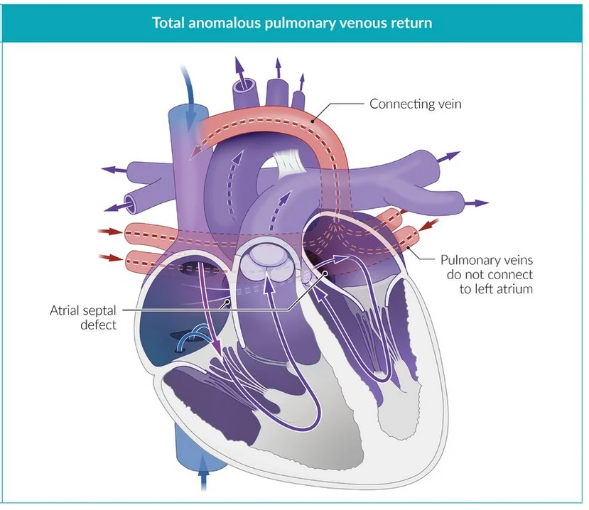
What this shows
Total anomalous pulmonary venous return
all four pulmonary veins drain into the systemic venous circulation (superior vena cava, coronary sinus, right atrium, or below the diaphragm into the portal vein) instead of the left atrium.- Oxygenated and deoxygenated blood mix completely in the right heart.
An atrial communication is obligatory
without return to the left heart there is no systemic output, so an atrial septal defect or foramen is needed for survival.Obstructed vs unobstructed
obstructed drainage (usually the infracardiac route through the liver) causes pulmonary venous hypertension and edema within hours, a surgical emergency.- Unobstructed drainage behaves like a giant atrial septal defect with cyanosis.
Clinical features and diagnosis
Cyanosis with respiratory distress
tachypnea and failure to thrive in the first week.Congestion
pulmonary venous congestion and right-sided heart failure (prominent neck veins, hepatomegaly).Examination
right ventricular heave, a widely split S2 with a loud P2, a soft pulmonary flow murmur; no murmur specific to the anomaly.Obstructed
profound cyanosis, severe distress and shock within hours of birth, easily mistaken for lung disease or persistent pulmonary hypertension.Transthoracic echocardiography is confirmatory
no pulmonary veins enter the left atrium, a venous confluence drains to a systemic vein, a dilated right heart with right-to-left atrial flow, and the site of obstruction.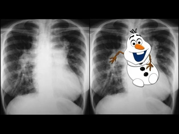
What this shows
Chest film
the "snowman" (figure-of-eight) sign with increased pulmonary vascular markings.- The dilated vertical vein and superior vena cava form the head, the heart the body.
- In obstruction: a normal-sized heart with diffuse pulmonary edema.
Electrocardiogram
right ventricular hypertrophy.| Obstructed (usually infracardiac) | Unobstructed (usually supracardiac) | |
|---|---|---|
| Onset | First hours: severe cyanosis, distress, shock | Weeks to months: mild cyanosis, failure to thrive, right heart failure |
| Chest film | Normal-sized heart, diffuse pulmonary edema ("white lungs") | Cardiomegaly, plethoric lungs, snowman |
| Mimics | Respiratory distress syndrome, persistent pulmonary hypertension, sepsis | Large atrial septal defect with cyanosis |
| Prostaglandin E1 | No benefit (may worsen the flooding) | Not needed |
| Surgery | Emergency | Within the first months |
Management and complications
Stabilization, then surgical repair
ventilation, correct acidosis, inotropes; then the venous confluence is anastomosed to the left atrium, the anomalous connection ligated and the atrial defect closed.- Emergency when obstructed.
In obstruction, avoid pulmonary vasodilators
they push more blood into a blocked pathway; balloon atrial septostomy for a restrictive atrial communication.Complications
untreated, death in infancy.- After repair, the main late problem is pulmonary vein stenosis at the anastomosis; also pulmonary hypertension and arrhythmias.
A 10-day-old has mild cyanosis, tachypnea, poor feeding and a right ventricular heave; the chest film shows a snowman-shaped heart with plethoric lungs. What is the lesion, and what makes it a surgical emergency?
Total anomalous pulmonary venous return (unobstructed, supracardiac: the snowman). It becomes an emergency when the drainage is obstructed (usually infracardiac): hours-old severe cyanosis and shock with a normal-sized heart and pulmonary edema, which needs emergency surgery.
How it's tested
A newborn with mild cyanosis at 2 weeks, tachypnea and hepatomegaly, an RV heave, a loud P2, plethoric lungs and a "snowman" cardiac silhouette: supracardiac TAPVR — echocardiogram and surgical repair.
A term newborn hours old with profound cyanosis, severe distress and a normal-sized heart with diffuse pulmonary edema; saturation fails to rise on 100% oxygen and does not improve with prostaglandin: obstructed (infracardiac) TAPVR — emergency surgery; nitric oxide and PGE1 do not help.
Why is an ASD obligatory in TAPVR: the left atrium receives no pulmonary venous blood directly — the only inflow to the systemic circulation is right-to-left across the atrial septum.
A repaired TAPVR patient at age 3 with progressive dyspnea and pulmonary hypertension: pulmonary vein stenosis at the anastomosis — the characteristic late complication.
Go deeper
Guidelines: 2018 AHA/ACC Adult Congenital Heart Disease Guideline
Related Step 2 pages: Approach to Neonatal Cyanosis, Approach to Congenital Heart Disease, Atrial Septal Defect, Persistent Pulmonary Hypertension of the Newborn, Transposition of the Great Arteries, Cardiac Anatomy
High-yield images2
You just read one page of 236
The rest of Step 2 CK, written exactly like this.
Rapid Review is the reading layer of a full Step 1 platform: a schedule fitted to your exam date, flashcards for every page, and a mentor a message away.
Every Step 1 system
Written the same way: what to know, how it’s tested, where to go deeper.
A plan built to your exam date
Tell us when you sit, and the schedule fits the whole library and your question bank to it.
Flashcards for every page
Ready-made spaced-repetition decks linked to each topic, so nothing you read gets forgotten.
