
Rapid Review·Cardiovascular
Select any text to highlight it or add a note.
PATHOLOGY
T1Must knowAortic Aneurysm & Dissection
FA P304-305, 319
Videos
2 to play here
Play 2 here
Focus on
The wall that dilates and the wall that splits, and the risk factor that separates the thoracic aorta from the abdominal.
Key takeaways
Aortic aneurysm
What it is
a localized pathologic dilation of the aorta. Abdominal or back pain signals leaking, dissection, or imminent rupture. The segments and their typical aneurysm sites are shown on the aortic segments diagram.Thoracic aortic aneurysm
- Associated with cystic medial degeneration: collagen, elastin and smooth muscle of the media are replaced by mucoid extracellular matrix. In Marfan the elastic lamellae fragment and are lost, with fibrosis, giving a "basket weave" pattern.
- Risk factors: hypertension, bicuspid aortic valve, connective tissue disease (Marfan), and tertiary syphilis, which causes obliterative endarteritis of the vasa vasorum.
- Aortic root dilatation causes aortic regurgitation. A large aneurysm also compresses its neighbours: hoarseness (recurrent laryngeal nerve), dysphagia (esophagus), cough (trachea).
Abdominal aortic aneurysm
- Atherosclerosis acts as a diffusion barrier to the media, which atrophies and weakens, with transmural inflammation through all three layers and extracellular matrix degradation.
- Risk factors: tobacco smoking (strongest), increasing age, male sex, family history.
- Palpable pulsatile abdominal mass that grows with time. Rupture, usually once it passes 5 cm, gives the rupture triad: pulsatile abdominal mass, acute abdominal or back pain, and resistant hypotension.
- Most often infrarenal, where the vasa vasorum supply is sparsest.
| Feature | Thoracic | Abdominal |
|---|---|---|
| Pathology | Cystic medial degeneration | Transmural inflammation with matrix degradation |
| Risk factors | Hypertension, bicuspid aortic valve, Marfan, tertiary syphilis | Smoking (strongest), age, male sex, family history |
| Location | Ascending aorta and arch | Most often infrarenal |
| Complication | Aortic root dilation causing aortic regurgitation | Rupture triad: pulsatile mass, pain, resistant hypotension |
| Mehlman's exam finding | "Visible pulsation above the manubrium", tracheal shift | "Visible pulsation in the epigastrium" |
Syphilitic heart disease
tertiary syphilis destroys the vasa vasorum of the aorta, so the wall atrophies and the aorta and valve ring dilate. Calcification of the aortic root and ascending arch gives the "tree bark" appearance. Causes ascending aortic aneurysm and aortic insufficiency.Aortic dissection
What it is
a longitudinal intimal tear creating a false lumen, visible as the intimal flap on the dissection on CT.Risk factors
hypertension (strongest risk factor), bicuspid aortic valve, and inherited connective tissue disease such as Marfan (through cystic medial degeneration).Presentation
tearing, sudden-onset chest pain radiating to the back, with markedly unequal arm blood pressures (more than 10 mm Hg). CXR shows mediastinal widening.Complications
organ ischemia when a branch artery is obstructed (coronary, renal), embolic stroke, aortic rupture, and pericardial tamponade, the most common cause of death.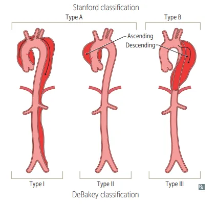
What this shows
The Stanford classification
drawn beside DeBakey's on the figure.Stanford type A (proximal)
involves the Ascending aorta, starting at the sinotubular junction and possibly extending into the arch or descending aorta. May cause acute aortic regurgitation or cardiac tamponade. Treatment is surgery.Stanford type B (distal)
involves only the descending aorta, Below the left subclavian. Treatment is beta-blockers, then vasodilators.| Feature | Type A (proximal) | Type B (distal) |
|---|---|---|
| Involves | Ascending aorta | Only descending, Below the left subclavian |
| Complications | Acute aortic regurgitation, cardiac tamponade | Organ ischemia |
| Treatment | Emergency surgery | Beta-blockers, then vasodilators |
| Mehlman's version | Labetalol plus surgery | Labetalol alone initially |
| Cause | Frequency | Distinguishing feature |
|---|---|---|
| Aortic dissection | About 3 in 4 | Tearing chest pain radiating to the back, widened mediastinum |
| Subclavian steal syndrome | About 1 in 4 | Dizziness on exerting the arm, no chest pain |
Traumatic aortic rupture
What it is
rupture from trauma or a deceleration injury such as a motor vehicle accident or significant fall.Where
most commonly at the aortic isthmus, the proximal descending aorta just distal to the left subclavian origin.Findings
a widened mediastinum on x-ray, a pulse discrepancy between the upper and lower extremities (as in coarctation), and an interscapular murmur from flow hitting the injured wall.Tearing chest pain to the back, arm pressures 40 mm Hg apart, and a blood pressure of 95/60. First drug?
Labetalol, even at that low pressure: the aim is lowering shear (dP/dt) on the torn wall, and a vasodilator first would raise it through reflex tachycardia.
How it's tested
Tearing chest pain radiating to the back with unequal arm blood pressures is aortic dissection, and labetalol is the answer even when the pressure is already low, because the goal is reducing shear rather than treating a number. That counterintuitive point is exactly what the question tests. Mehlman's prior is useful: when arm pressures differ, dissection is about three times more likely than the alternatives.
For the aneurysms, split them by risk factor. Abdominal is smoking and atherosclerosis, screened with a one-time ultrasound in men 65 and over who ever smoked; thoracic is cystic medial degeneration, from Marfan, a bicuspid valve, or tertiary syphilis, which obliterates the vasa vasorum and gives the tree-bark aorta with aortic regurgitation. Mehlman's counterintuitive addition is that diabetes protects against aneurysm formation, since glycosylation stiffens the wall rather than weakening it.
Go deeper
First Aid 2026 — CV/Pathology (p.304-305, 319) · B&B — Aneurysms; aortic dissection · Mehlman — HY Cardio (AAA screening and repair thresholds, Stanford A/B, labetalol first)
High-yield images4
Flashcards for this page
Card 1 of 5 · try-out only, nothing is saved
(...) sex
Increased (...)
(...)
(...)
You just read one page of 944
The rest of Step 1, written exactly like this.
Rapid Review is the reading layer of a full Step 1 platform: a schedule fitted to your exam date, flashcards for every page, and a mentor a message away.
Every Step 1 system
Written the same way: what to know, how it’s tested, where to go deeper.
A plan built to your exam date
Tell us when you sit, and the schedule fits the whole library and your question bank to it.
Flashcards for every page
Ready-made spaced-repetition decks linked to each topic, so nothing you read gets forgotten.
