
Rapid Review·Cardiovascular
Select any text to highlight it or add a note.
PATHOLOGY
T1Must knowAtherosclerosis, Arteriolosclerosis & Cholesterol Emboli
FA P303-304
Videos
1 to play here
Play 1 here
Focus on
The skin findings that betray hyperlipidemia, the cascade that builds a plaque, and the two ways small vessels thicken.
Key takeaways
The signs that betray hyperlipidemia
Xanthomas
plaques or nodules of lipid-laden histiocytes in skin, classically the eyelids, where they are called xanthelasma.Tendinous xanthoma
lipid deposited in tendon, especially the Achilles and the finger extensors. Strongly associated with familial hypercholesterolemia.Corneal arcus
lipid in the cornea. Common and benign in older adults (arcus senilis), but early onset means hypercholesterolemia.| Sign | Location | Association |
|---|---|---|
| Xanthoma / xanthelasma | Skin, especially the eyelids | Lipid-laden histiocytes |
| Tendinous xanthoma | Achilles tendon, finger extensors | Familial hypercholesterolemia |
| Corneal arcus | Cornea | Normal in older adults; early onset means hypercholesterolemia |
Atherosclerosis
What it is
the common form of arteriosclerosis (hard arteries: a thickened wall that has lost its elasticity), affecting elastic and large-to-medium muscular arteries. It is driven by cholesterol plaque deposition in the tunica intima, causing narrowing and inflammation. May be asymptomatic, or present as angina or claudication.Location, in descending order of involvement
abdominal aorta > coronary artery > popliteal artery > carotid artery > circle of Willis. "A copy cat named Willis."Risk factors
- Modifiable: hypertension, tobacco smoking, dyslipidemia (high LDL, low HDL), diabetes.
- Non-modifiable: age, male sex, postmenopausal status, family history.
How a plaque is built
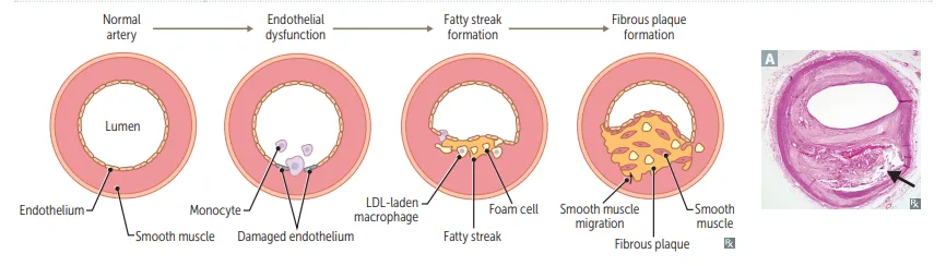
What this shows
Endothelial cell dysfunction
inflammation is central from here on.Macrophage and LDL accumulation.
Foam cell formation
macrophages engulf oxidized LDL.Fatty streaks
the earliest visible lesion.Smooth muscle migration, proliferation, and matrix deposition
platelets adhere to the abnormal endothelium and, with the macrophages, release PDGF and FGF, which build the fibrous cap of the mature atheroma.Fibrous plaque, then complex atheroma, then calcification
calcification proteins such as osteopontin let fibroblasts behave like osteoblasts, and the calcium content correlates with complication risk.Why plaques rupture
stability depends on the mechanical strength of the fibrous cap, and intimal macrophages weaken it by secreting metalloproteinases that digest the matrix.Statins lower LDL
which slows plaque progression and reduces the risk of plaque rupture.Complications
ischemia, infarction, aneurysm formation, peripheral vascular disease, thrombosis, embolism, renovascular hypertension, and subclavian steal syndrome.| Step | Event | Key mediators |
|---|---|---|
| 1 | Endothelial cell dysfunction | Shear stress, smoking, hypertension |
| 2 | Macrophage and LDL accumulation | Adhesion molecules |
| 3 | Foam cell formation | Macrophages engulf oxidized LDL |
| 4 | Fatty streaks | The earliest visible lesion |
| 5 | Smooth muscle migration, proliferation, matrix deposition | PDGF and FGF |
| 6 | Fibrous plaque, complex atheroma, calcification | Calcium correlates with complication risk |
The other two arterioscleroses
| Type | Vessels | Pathology |
|---|---|---|
| Atherosclerosis | Elastic and large-to-medium muscular arteries | Intimal cholesterol plaque |
| Arteriolosclerosis | Small arteries and arterioles | Hyaline or hyperplastic |
| Mönckeberg (medial calcific sclerosis) | Medium arteries | Calcification of the media, and it does not obstruct the lumen |
Arteriolosclerosis
affects small arteries and arterioles, in two forms:- Hyaline: wall thickening from plasma protein leak into the subendothelium. Seen in hypertension or diabetes.
- Hyperplastic: "onion skinning" from smooth muscle proliferation. Seen in severe (malignant) hypertension.
Mönckeberg sclerosis
dystrophic calcification of the tunica media and internal elastic lamina of medium-sized arteries. The intima is spared, so flow is not obstructed; x-ray shows a "pipestem" vessel.| Feature | Hyaline | Hyperplastic |
|---|---|---|
| Mechanism | Plasma protein leak into subendothelium | Smooth muscle proliferation |
| Appearance | Homogeneous pink wall thickening | "Onion skinning" |
| Setting | Hypertension or diabetes mellitus | Severe (malignant) hypertension |
Cholesterol emboli syndrome
What it is
microembolization of cholesterol crystals dislodged from atherosclerotic plaques in large arteries, usually the aorta, causing end-organ damage plus an inflammatory response.Findings
livedo reticularis, digital ischemia (blue toe syndrome), acute renal failure, stroke, gut ischemia.The key clue is that pulses remain palpable
only the small vessels are showered while the large arteries stay patent.Trigger
often follows an invasive vascular procedure: angiography, angioplasty, endovascular grafting.| Feature | Cholesterol emboli syndrome | Large-artery occlusion |
|---|---|---|
| Pulses | PALPABLE, large arteries unaffected | Absent |
| Findings | Livedo reticularis, blue toe syndrome, acute kidney injury, stroke | Distal ischemia of a whole territory |
| Trigger | After angiography, angioplasty, or grafting | Thrombus or embolus |
Two days after cardiac catheterization a patient has blue toes and a mottled leg, yet the pedal pulses are normal. What happened?
Cholesterol emboli syndrome: the procedure showered plaque crystals into the small vessels, and the preserved pulses are the diagnostic key.
How it's tested
Foam cells are macrophages that have engulfed oxidized LDL, and they are the cellular signature of the fatty streak, the earliest visible lesion. A stem asking what the earliest change is wants endothelial dysfunction, and one asking what the earliest visible lesion is wants the fatty streak, so keep those two apart. The location hierarchy is worth carrying as a single unit: abdominal aorta, then coronary, popliteal, carotid, and circle of Willis. Mönckeberg is the distractor that does not narrow the lumen.
Blue toes with intact pedal pulses after a recent cardiac catheterization is cholesterol emboli syndrome, and the preserved pulses are the diagnostic key, because only the small vessels are showered. The arteriolosclerosis pair is decided by the setting: hyaline thickening goes with long-standing hypertension and diabetes, whereas hyperplastic, the onion-skin change, goes with malignant hypertension.
Go deeper
First Aid 2026 — CV/Pathology (p.303-304) · B&B — Atherosclerosis; arteriolosclerosis · Mehlman — HY Cardio (plaque steps, Mönckeberg); familial dyslipidemias cross-ref Biochemistry
High-yield images1
Flashcards for this page
Card 1 of 5 · try-out only, nothing is saved
(...)-sized arteries
You just read one page of 944
The rest of Step 1, written exactly like this.
Rapid Review is the reading layer of a full Step 1 platform: a schedule fitted to your exam date, flashcards for every page, and a mentor a message away.
Every Step 1 system
Written the same way: what to know, how it’s tested, where to go deeper.
A plan built to your exam date
Tell us when you sit, and the schedule fits the whole library and your question bank to it.
Flashcards for every page
Ready-made spaced-repetition decks linked to each topic, so nothing you read gets forgotten.
