
Rapid Review·Cardiovascular
Select any text to highlight it or add a note.
PATHOLOGY
T2High yieldCardiomyopathies & Myocarditis
FA P314-315, 320
Videos
1 to play here
Play 1 here
Focus on
Three cardiomyopathies, three geometries, and the inflammation that turns into the commonest of them.
Key takeaways
The sarcomere logic
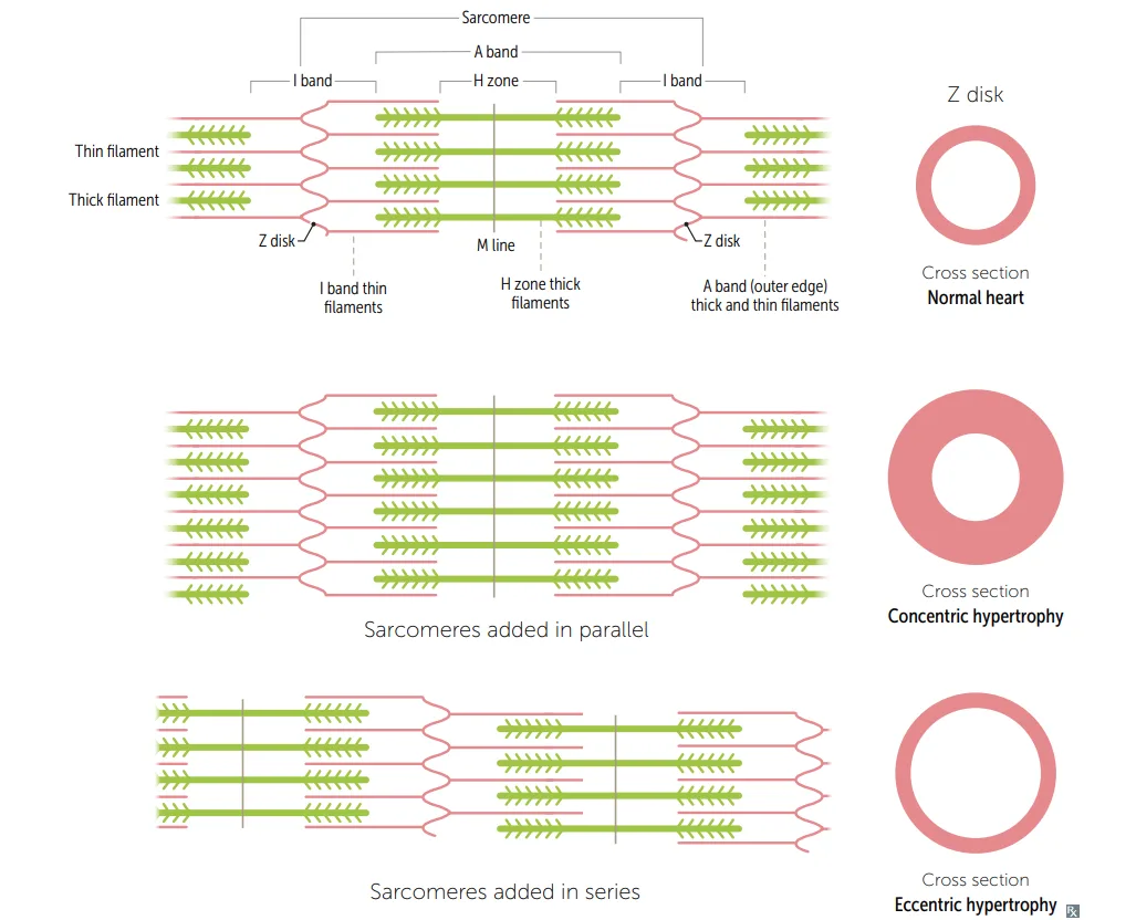
What this shows
Sarcomeres added in parallel thicken the wall inward
concentric hypertrophy, the pressure-overload pattern.Sarcomeres added in series lengthen the fibre
eccentric hypertrophy, the volume-overload pattern with a dilated, thin-walled chamber.The mutation mnemonic
"Titin is thin, myosin is more": titin gives dilated, myosin gives hypertrophic.Dilated cardiomyopathy
How common
the most common, about 90% of cases. Often idiopathic or familial, from a TTN mutation shortening titin.Etiologies
drugs (alcohol, cocaine, doxorubicin), infection (coxsackie B, Chagas, so ask where the patient has lived), ischemia (CAD), systemic disease (hemochromatosis, sarcoidosis, thyrotoxicosis, wet beriberi), and peripartum cardiomyopathy, during or shortly after pregnancy. In Chagas the usual cause of death is sudden cardiac death from ventricular arrhythmia.Findings
heart failure, S3, systolic regurgitant murmur, dilated heart on echo (large LV cavity, low EF, normal or thin wall), balloon appearance on CXR. Systolic dysfunction with eccentric hypertrophy.Contrast an athlete's heart
where balanced enlargement physiologically supports a higher stroke volume.Treatment
sodium restriction, ACE inhibitors or ARBs, beta-blockers, sacubitril/valsartan, diuretics, spironolactone, ICD, transplant.Hypertrophic cardiomyopathy
How common
the second most common cardiomyopathy, and the most common cause of sudden cardiac death in athletes and teenagers. 60 to 70% familial, autosomal dominant, from sarcomeric mutations in beta-myosin heavy chain or myosin-binding protein C. Also from chronic hypertension and Friedreich ataxia.Presentation
syncope during exercise, and a risk of sudden death in young athletes from ventricular arrhythmia.Findings
S4, systolic murmur, sometimes mitral regurgitation from impaired mitral closure. Diastolic dysfunction with concentric hypertrophy, often septal; on echo a small LV cavity with a normal EF and a thick wall. Histology shows myofibrillar disarray and fibrosis.HOCM
the subset where the LV outflow tract is obstructed by asymmetric septal hypertrophy plus systolic anterior motion of the mitral valve, with a bifid carotid pulse.Treatment
the aim is to keep the LV full (high preload, low heart rate, low contractility). Beta-blockers or a nondihydropyridine calcium channel blocker (verapamil) lengthen diastolic filling and raise end-systolic volume; mavacamten if refractory; stop high-intensity athletics; ICD if high risk. AVOID anything that lowers preload (diuretics, vasodilators), because a smaller ventricle worsens the obstruction.| Intervention | Dilated and most lesions | Hypertrophic |
|---|---|---|
| Lower preload (diuretics, vasodilators, Valsalva) | Improves congestion | WORSENS obstruction, contraindicated |
| Raise preload (squatting, leg raise) | Increases congestion | Relieves obstruction |
Restrictive (infiltrative) cardiomyopathy
Causes, by "PLEASe Help"
- Postradiation fibrosis
- Löffler endocarditis, from hypereosinophilic syndrome with eosinophilic myocardial infiltrates
- Endocardial fibroelastosis, thick fibroelastic endocardium in young children
- Amyloidosis
- Sarcoidosis
- Hemochromatosis
Findings
diastolic dysfunction from reduced compliance; echo shows a normal cavity, a normal or thick wall, and a preserved EF. The giveaway is a low-voltage ECG despite a thick myocardium, classically in amyloidosis.| Feature | Dilated | Hypertrophic | Restrictive |
|---|---|---|---|
| Frequency | Most common, 90% | 60 to 70% familial | Least |
| Dysfunction | Systolic | Diastolic | Diastolic |
| Geometry | Eccentric, sarcomeres in series | Concentric, sarcomeres in parallel | Normal thickness or infiltrated |
| Heart sound | S3 | S4 | Variable |
| Genetics | TTN (titin) | Beta-myosin heavy chain, myosin-binding protein C | Depends on cause |
| EF | Reduced | Normal or raised | Normal |
| ECG | Non-specific | LVH | Low voltage despite thick myocardium |
| Treatment | ACEi/ARB, beta-blockers, spironolactone, ICD | Beta-blockers, non-DHP CCB, mavacamten; avoid preload reduction | Treat the underlying disease |
| Category | Causes |
|---|---|
| Drugs | Alcohol, cocaine, doxorubicin |
| Infection | Coxsackie B, Chagas disease |
| Ischemia | Coronary artery disease |
| Systemic | Hemochromatosis, sarcoidosis, thyrotoxicosis, wet beriberi |
| Peripartum | Peripartum cardiomyopathy |
| Feature | Dilated | Hypertrophic | Restrictive | Athlete's heart |
|---|---|---|---|---|
| LV cavity | Large | Small | Normal | Normal or large |
| Diastolic function | Normal | Impaired | Impaired | Normal |
| Wall thickness | Normal or thin | Thick | Normal or thick | Variable |
| Contractility | Reduced | Normal or raised | Normal | Raised |
| LV mass | Raised | Raised | Normal | Raised |
| EF | Reduced | Preserved | Preserved | Preserved |
Athlete's heart
What it is
the cardiac changes of athletic training, which differ by the kind of training.| Feature | Strength training | Endurance training |
|---|---|---|
| Mechanism | Muscle contraction squeezes the systemic arteries, raising afterload | Skeletal muscle vasodilation lowers SVR while sympathetic venoconstriction raises preload, so stroke volume rises with an unchanged EF |
| Left ventricle | Concentric hypertrophy (thicker wall) | Eccentric hypertrophy (bigger cavity) |
| Right ventricle | No change | Larger cavity (a pulmonic flow murmur from the extra RV stroke volume) |
| Diastolic filling | No change | Increased |
| EF | Unchanged, or slightly raised | Unchanged |
Myocarditis
What it is
inflammation of the myocardium, and a major cause of sudden cardiac death in adults under 40.Presentation is highly variable
dyspnea, chest pain, fever, arrhythmias. Persistent tachycardia out of proportion to the fever is characteristic. A young patient who develops heart failure after a viral prodrome (fever, congestion, myalgias) has viral myocarditis until proven otherwise.Complications
sudden death, arrhythmias, heart block, dilated cardiomyopathy, heart failure, mural thrombus with systemic emboli.| Category | Agents |
|---|---|
| Viral | Coxsackie B, adenovirus, parvovirus B19, HIV, HHV-6, COVID-19. Lymphocytic infiltrate with focal necrosis is highly indicative |
| Parasitic | Trypanosoma cruzi (Chagas), Toxoplasma gondii |
| Bacterial | Borrelia burgdorferi, Mycoplasma, C diphtheriae |
| Toxins | Carbon monoxide, black widow venom |
| Drugs | Doxorubicin, cocaine |
| Autoimmune | Kawasaki, sarcoidosis, SLE, polymyositis and dermatomyositis, rheumatic fever |
A thick-walled heart with a low-voltage ECG. Which cardiomyopathy, and which cause?
Restrictive, from amyloidosis: the infiltrate adds wall but not electrically active muscle.
How it's tested
A young athlete with syncope during exercise and a systolic murmur that gets louder on standing has hypertrophic cardiomyopathy, and the management point that carries the question is that diuretics and vasodilators are contraindicated, because reducing preload shrinks the cavity and worsens the outflow obstruction. A low-voltage ECG in a thick-walled heart is the amyloidosis giveaway, since infiltrate is not electrically active muscle. Myocarditis is tested as the young patient who dies suddenly or presents in new heart failure after a viral prodrome, with coxsackie B the organism to name, and it belongs here because it is a leading route into dilated cardiomyopathy.
Go deeper
First Aid 2026 — CV/Pathology (p.314-315, 320) · B&B — Dilated, hypertrophic & restrictive cardiomyopathy; myocarditis · Mehlman — HY Cardio (cardiomyopathy causes, HOCM management)
High-yield images1
Flashcards for this page
Card 1 of 5 · try-out only, nothing is saved
(...) endocarditis
You just read one page of 944
The rest of Step 1, written exactly like this.
Rapid Review is the reading layer of a full Step 1 platform: a schedule fitted to your exam date, flashcards for every page, and a mentor a message away.
Every Step 1 system
Written the same way: what to know, how it’s tested, where to go deeper.
A plan built to your exam date
Tell us when you sit, and the schedule fits the whole library and your question bank to it.
Flashcards for every page
Ready-made spaced-repetition decks linked to each topic, so nothing you read gets forgotten.
