
Rapid Review·Cardiovascular
Select any text to highlight it or add a note.
PHYSIOLOGY
T1Must knowElectrocardiogram
P297
Videos
1 to play here
Play 1 here
Focus on
The conduction pathway, the intervals, and what each wave means.
Key takeaways
The conduction pathway
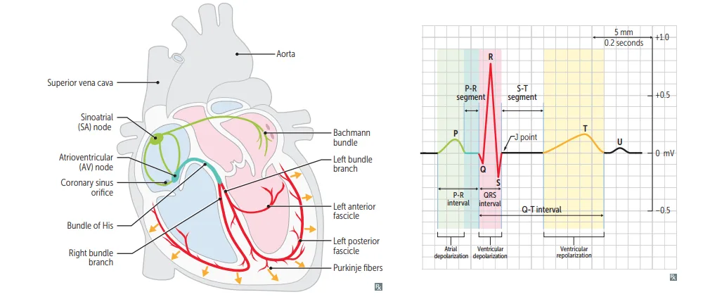
What this shows
The route, traced on the left half of the figure
SA node to the right atrium (internodal tracts) and left atrium (Bachmann bundle), then AV node, bundle of His, the right and left bundle branches (the left dividing into anterior and posterior fascicles), then Purkinje fibers to the ventricles.SA node
sits in the upper crista terminalis near the SVC, and is the dominant pacemaker.AV node
sits in the interatrial septum near the coronary sinus opening, supplied by the PDA. Its 100 msec delay allows ventricular filling.| Ranking | Order |
|---|---|
| Pacemaker rate (intrinsic firing) | SA > atria > AV > bundle of His, Purkinje, ventricles |
| Conduction speed | His-Purkinje > Atria > Ventricles > AV node (He Parks At Ventura AVenue) |
Waves and intervals
Labelled on the right half of the same figure
- P wave: atrial depolarization.
- PR interval: normally 120 to 200 msec.
- QRS complex: normally 100 msec or less.
- QT interval: ventricular depolarization, contraction, and repolarization together.
- T wave: ventricular repolarization. Inversion may mean ischemia or recent MI.
- J point: the junction between the end of QRS and the start of the ST segment.
- ST segment: isoelectric, with the ventricles fully depolarized.
- U wave: prominent in hypokalemia (think hypUkalemia) and in bradycardia.
| Component | Represents | Normal value |
|---|---|---|
| P wave | Atrial depolarization | |
| PR interval | Start of atrial to start of ventricular depolarization | 120 to 200 msec |
| QRS complex | Ventricular depolarization | 100 msec or less |
| QT interval | Ventricular depolarization, contraction and repolarization | |
| T wave | Ventricular repolarization | Inversion suggests ischemia or recent MI |
| ST segment | Ventricles depolarized (isoelectric) | |
| U wave | Late repolarization | Prominent in hypokalemia and bradycardia |
An inferior MI presents with bradycardia and heart block. Which artery, and which structure did it starve?
The posterior descending artery, which supplies the AV node.
How it's tested
The AV node conducts most slowly of all, and that roughly 100 millisecond delay is a feature rather than a defect, because it gives the ventricles time to fill before they contract. Its blood supply comes from the posterior descending artery, which is why an inferior MI can present with bradycardia or heart block. The other easy point is the U wave, which is the ECG signature of hypokalemia.
Go deeper
First Aid 2026 — CV/Physiology (p.297) · B&B — ECG basics · Mehlman — HY Cardio (intervals, U wave and hypokalemia)
High-yield images1
Flashcards for this page
Card 1 of 5 · try-out only, nothing is saved
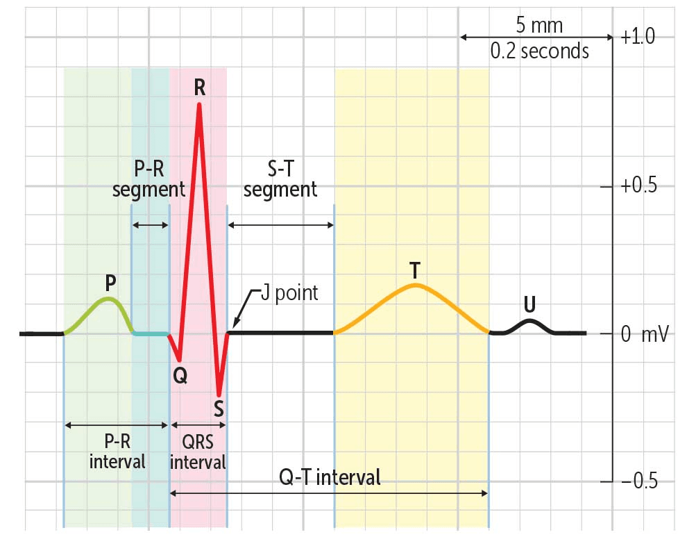
Time from the beginning of (...) to the beginning of (...)
You just read one page of 944
The rest of Step 1, written exactly like this.
Rapid Review is the reading layer of a full Step 1 platform: a schedule fitted to your exam date, flashcards for every page, and a mentor a message away.
Every Step 1 system
Written the same way: what to know, how it’s tested, where to go deeper.
A plan built to your exam date
Tell us when you sit, and the schedule fits the whole library and your question bank to it.
Flashcards for every page
Ready-made spaced-repetition decks linked to each topic, so nothing you read gets forgotten.
