
Rapid Review·Cardiovascular
Select any text to highlight it or add a note.
ANATOMY
T2High yieldHeart Anatomy
FA P287
Videos
2 to play here
Play 2 here
Focus on
Which chamber sits where, because position explains dysphagia, hoarseness, trauma, and every coronary infarct pattern.
Key takeaways
Position, and what each position causes
The heart sits in the MIDDLE mediastinum
which is why a pericardial effusion widens the cardiac silhouette centrally and why the phrenic nerves run beside it.LA is the most POSTERIOR chamber
enlargement, classically from mitral stenosis, compresses the esophagus (dysphagia) and the left recurrent laryngeal nerve (hoarseness, Ortner syndrome).- The same relationship is what makes transesophageal echocardiography so useful: looking anteriorly from the esophagus sees the left atrium, and looking posteriorly sees the descending aorta.
RV is the most ANTERIOR chamber
so it forms the anterior border and is the chamber most commonly injured in penetrating trauma, such as a stab wound.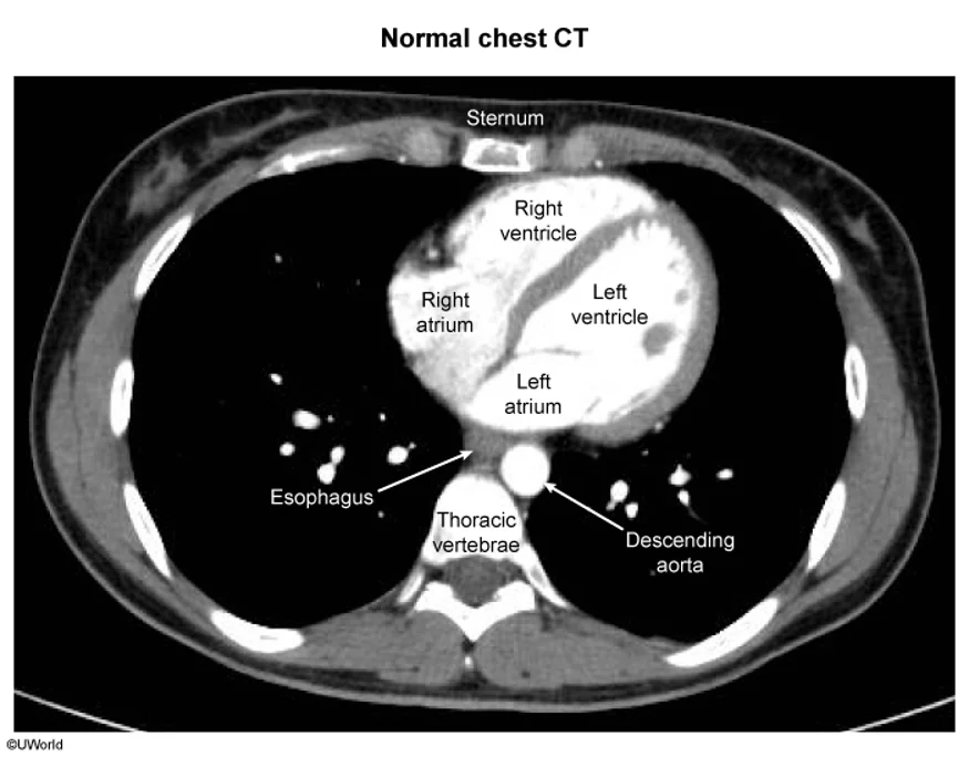
What this shows
On the frontal view the borders are formed by different chambers again
the right border by the right atrium, the left border by the left atrium and left ventricle, and the inferior (diaphragmatic) surface is about two thirds LV and one third RV.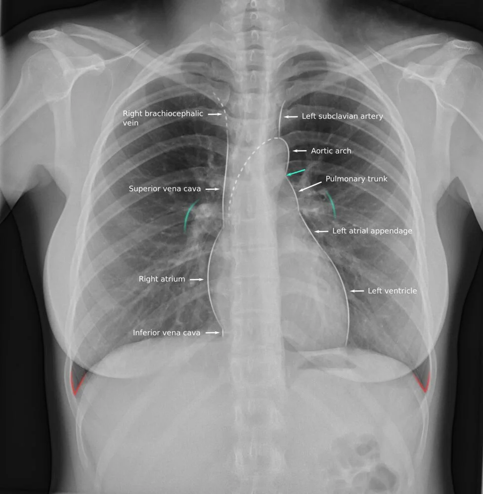
What this shows
| Chamber | Position | Clinical consequence |
|---|---|---|
| Left atrium | Most posterior | Enlargement gives dysphagia (esophagus) and hoarseness (left recurrent laryngeal nerve, Ortner syndrome) |
| Right ventricle | Most anterior | Most commonly injured in trauma |
| Right atrium | Forms the right border | Right heart border on chest film |
| Left ventricle | Forms the left border, 2/3 of the diaphragmatic surface | Left heart border, apex beat |
Right atrial landmarks
all visible on the right atrial anatomy: the SA node at the crista terminalis near the SVC, the AV node in the interatrial septum, the fossa ovalis, and the coronary sinus ostium.Why the right ventricle tolerates ischemia
The RV survives ischemia that would infarct the LV, for five reasons
all of them follow from the RV doing far less work against far lower pressure.| Feature | Left ventricle | Right ventricle |
|---|---|---|
| Muscle mass | High | Low |
| Resting oxygen extraction | High, near maximal | Low, so there is reserve |
| Coronary perfusion | During diastole only | Throughout the cardiac cycle |
| Collateral circulation | Less developed | More developed |
| Ischemic preconditioning | Low | High |
The LV is perfused only in diastole
in systole its wall squeezes its own vessels shut, whereas the RV generates far lower pressure and never occludes them, so it is perfused continuously. That single difference is also why tachycardia, which shortens diastole, provokes LV ischemia specifically.Pericardium
What this shows
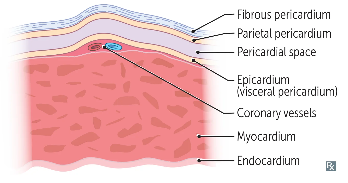
The layers from outer to inner
fibrous pericardium, then parietal pericardium, then the pericardial space, then the epicardium (visceral pericardium), which carries the coronary vessels.The pericardium is innervated by the PHRENIC nerve
which arises from C3 to C5, and that is why pericarditis refers pain to the neck, shoulders or arms, most often the left. See Pericardial Disease: Pericarditis, Tamponade & Constriction.Coronary blood supply
Coronary blood flow to the LV and septum peaks in EARLY DIASTOLE
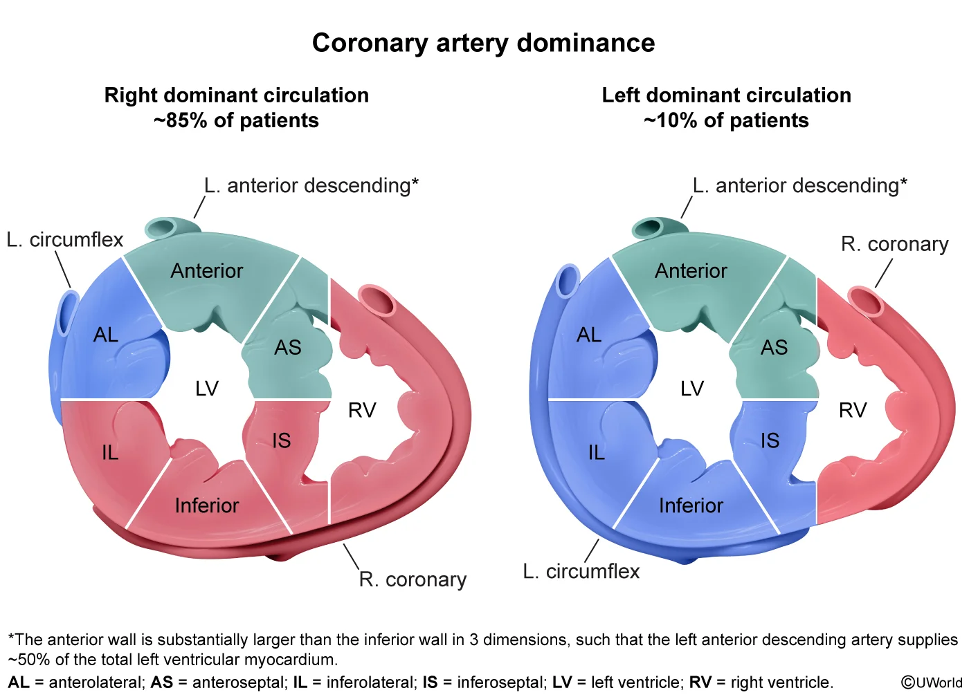
which is the mechanical reason tachycardia, by shortening diastole, provokes ischemia.Compare the anterior and the left-dominant posterior views
the anterior coronary view against the posterior (left-dominant) view, and the territory map on the coronary territories.- LAD supplies the anterior 2/3 of the interventricular septum, the anterolateral papillary muscle, and the anterior LV surface. Most commonly occluded.
- Left circumflex (LCX) supplies the lateral surface of the LV and also the anterolateral papillary muscle.
- RCA supplies the SA and AV nodes, and its right (acute) marginal branch supplies the RV.
- PDA supplies the posterior 1/3 of the interventricular septum, the posterior ventricular walls, the inferior wall of the LV, and the posteromedial papillary muscle.
An infarct of the AV nodal artery causes nodal dysfunction
bradycardia or heart block, which is counteracted by atropine. That is shown on the conduction system blood supply.The coronary sinus runs in the LEFT AV groove and drains into the RA
it carries the most deoxygenated blood in the body, because the myocardium extracts oxygen near-maximally at rest, and it dilates with anything that raises right atrial pressure. See the coronary sinus.| Artery | Supplies | Clinical note |
|---|---|---|
| Left anterior descending (LAD) | Anterior LV surface, anterior 2/3 of septum, anterolateral papillary muscle | Most commonly occluded artery |
| Left circumflex (LCX) | Lateral LV surface, anterolateral papillary muscle | Lateral infarcts |
| Right coronary (RCA) | SA and AV nodes; right marginal branch supplies the RV | AV nodal infarct gives bradycardia or heart block, counteracted by atropine |
| Posterior descending (PDA) | Inferior wall of LV, posterior 1/3 of septum, posteromedial papillary muscle | Inferior infarcts |
Coronary dominance
What this shows
Dominance is defined by which artery gives the PDA
- Right-dominant, the most common: the PDA arises from the RCA.
- Left-dominant: the PDA arises from the LCX.
- Codominant: the PDA arises from both the LCX and the RCA.
The AV nodal artery arises from whichever artery is dominant
from the LCX in left dominance, and from the RCA in right-dominant and codominant hearts. That is the rule behind every "which artery, and what happened to the AV node" question.| Pattern | PDA arises from | Frequency | AV nodal artery from |
|---|---|---|---|
| Right-dominant | RCA | Most common, about 85% | RCA |
| Left-dominant | LCX | About 5 to 10% | LCX |
| Codominant | Both LCX and RCA | About 10 to 20% | RCA |
| Papillary muscle | Blood supply | Rupture risk |
|---|---|---|
| Posteromedial | Single, PDA only | Much higher, the classic post-MI rupture |
| Anterolateral | Dual, LAD and LCX | Lower |
Conduction pathway
laid out on the cardiac conduction system: SA node to AV node to bundle of His to the right and left bundle branches to the Purkinje network.The great vessels
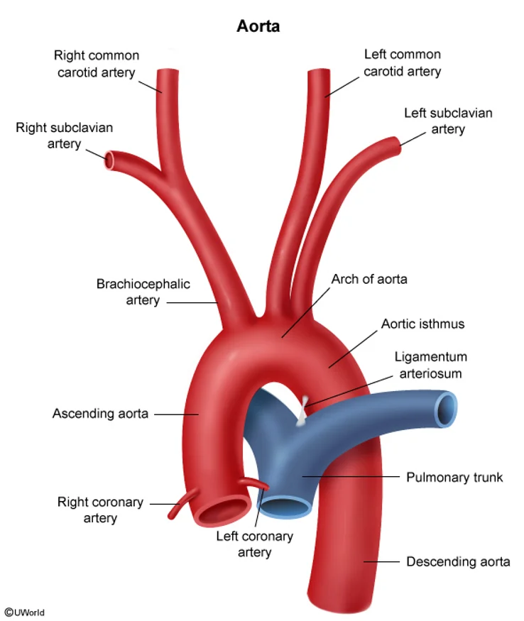
What this shows
The aorta has three segments, and each supplies a different region
- Ascending aorta: gives the right and left coronary arteries, supplying the heart itself and nothing else.
- Aortic arch: supplies the upper limbs, head and neck through three branches in order, the brachiocephalic artery (dividing into right subclavian and right common carotid), the left common carotid, and the left subclavian.
- Descending (thoracic) aorta: gives the bronchial arteries, contributing the second half of the lung's dual blood supply, the posterior intercostal arteries, whose enlargement causes rib notching in coarctation, and the esophageal arteries. It becomes the abdominal aorta at the aortic hiatus of the diaphragm, at T12.
The subclavian artery is the one behind subclavian steal
its branches are the thyrocervical trunk (giving the inferior thyroid artery), the vertebral artery, which joins its fellow to form the basilar artery supplying the brainstem and posterior circulation, and the anterior intercostal arteries. See Peripheral Artery Disease, Acute Limb Ischemia & Subclavian Steal.- It continues as the axillary then brachial artery, which accompanies the median nerve and is injured in supracondylar humeral fractures; its deep brachial branch accompanies the radial nerve and is injured in midshaft humeral fractures.
The common carotid divides into internal and external
- Internal carotid supplies mainly intracranial structures, giving the ophthalmic artery (and its central retinal branch), the anterior cerebral and the middle cerebral arteries.
- External carotid supplies mainly extracranial structures, giving the superior thyroid and maxillary arteries, the latter giving the middle meningeal artery torn in epidural hemorrhage.
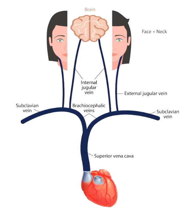
What this shows
The SVC drains the whole upper body, so its obstruction is BILATERAL
it receives the right and left brachiocephalic veins and returns blood to the right atrium. SVC syndrome gives bilateral swelling of the head, neck and both upper limbs.A brachiocephalic vein obstruction is UNILATERAL
because each brachiocephalic vein drains only its own side, receiving that side's subclavian and internal jugular veins. The external jugular vein, draining the face and neck, joins the subclavian.- The summary worth carrying: brain to internal jugular to brachiocephalic to SVC, and face and neck to external jugular to subclavian to brachiocephalic to SVC.
The azygos system is the collateral route between SVC and IVC
the azygos vein rises from the ascending lumbar veins along the right side of the vertebral column and connects the SVC to the IVC, so it becomes an alternative path back to the right atrium if either cava is obstructed. The hemiazygos runs up the left side, from the left renal vein below to the azygos above.A 64-year-old man has ST elevation in leads II, III and aVF, and develops a heart rate of 38 with complete heart block. Angiography shows occlusion of a vessel that also gives rise to the posterior descending artery. Three days later he becomes acutely dyspneic with a new holosystolic murmur at the apex. Name the artery, the dominance pattern, the reason for the bradycardia, and the day-3 complication.
The artery is the right coronary artery and the pattern is right-dominant, the arrangement in roughly 85% of people, established by the stem's statement that this vessel gives the PDA. The ECG confirms it: leads II, III and aVF view the inferior wall, which the PDA supplies. The bradycardia and complete heart block follow because the AV nodal artery arises from whichever artery is dominant, so in a right-dominant heart the RCA supplies both the SA and the AV node; this is why inferior infarcts, not anterior ones, present with nodal block, and the answer for management is atropine. Had the patient been left-dominant, the same inferior infarct would have come from the left circumflex and the AV nodal artery would have arisen from the LCX instead. The day-3 complication is posteromedial papillary muscle rupture causing acute mitral regurgitation, and the anatomy explains why that muscle and not the other: the posteromedial papillary muscle has a single blood supply, from the PDA, while the anterolateral has a dual supply from the LAD and LCX and is therefore protected. The timing fits, since rupture classically occurs 3 to 5 days after infarction, when the necrotic tissue is at its weakest.
How it's tested
The posteromedial papillary muscle has a single blood supply from the posterior descending artery, while the anterolateral has a dual supply. That asymmetry is the entire reason post-MI papillary muscle rupture is posteromedial and produces acute mitral regurgitation, and it is worth deriving rather than memorising. The left atrium being most posterior explains both the dysphagia and the hoarseness of mitral stenosis. Separately, because the RCA supplies both the SA and AV nodes in most people, an inferior MI is the one that brings bradycardia and heart block.
Go deeper
First Aid 2026 — CV/Anatomy (p.287) · B&B — Coronary circulation · Mehlman — HY Cardio (coronary territories, inferior MI and bradycardia)
High-yield images13
Flashcards for this page
Card 1 of 5 · try-out only, nothing is saved
(...) nerve
You just read one page of 944
The rest of Step 1, written exactly like this.
Rapid Review is the reading layer of a full Step 1 platform: a schedule fitted to your exam date, flashcards for every page, and a mentor a message away.
Every Step 1 system
Written the same way: what to know, how it’s tested, where to go deeper.
A plan built to your exam date
Tell us when you sit, and the schedule fits the whole library and your question bank to it.
Flashcards for every page
Ready-made spaced-repetition decks linked to each topic, so nothing you read gets forgotten.
