
Rapid Review·Cardiovascular
Select any text to highlight it or add a note.
EMBRYOLOGY
T1Must knowFetal Circulation & Postnatal Derivatives
FA P286
Focus on
Three shunts that route blood around the liver and lungs, the breath that shuts them down, and the ligaments they leave behind.
Key takeaways
The three shunts
Fetal circulation is built to bypass two organs that are not working yet
the liver and the lungs. Each shunt exists to route blood around one of them.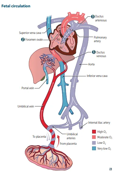
What this shows
Ductus venosus: umbilical vein into the IVC, bypassing the HEPATIC circulation
the umbilical vein carries the most oxygenated blood in the fetus, at about 80% saturation and a PO2 near 30 mmHg, and the placenta itself is the highest-oxygen point.Foramen ovale: IVC blood crosses from RA into LA, bypassing the right ventricle and lungs
so the best-oxygenated blood reaches the left ventricle, the ascending aorta, and therefore the head, neck and arms first.Ductus arteriosus: pulmonary artery into the descending aorta, bypassing the lungs
deoxygenated blood returning through the SVC goes RA to RV to pulmonary trunk, and is then diverted through the duct because fetal pulmonary vascular resistance is high.- That resistance is also why pressure on the right side of the fetal circulation is higher than on the left, the reverse of postnatal life, and it is what holds the foramen ovale open.
- From the descending aorta the blood reaches the common iliac, internal iliac and umbilical arteries back to the placenta, which carries the lowest oxygen saturation in the circuit.
| Shunt | Route | What it bypasses | Becomes |
|---|---|---|---|
| Ductus venosus | Umbilical vein to IVC | Hepatic circulation | Ligamentum venosum |
| Foramen ovale | IVC to RA to LA | Right ventricle and lungs | Fossa ovalis |
| Ductus arteriosus | Pulmonary artery to descending aorta | Lungs (high fetal PVR) | Ligamentum arteriosum |
What the first breath changes
Everything at birth follows from the lungs inflating
- The infant breathes, so pulmonary vascular resistance falls.
- LA pressure rises above RA pressure, so the foramen ovale closes, becoming the fossa ovalis.
- Arterial oxygen rises and, with placental separation, prostaglandins fall, so the ductus arteriosus closes.
| Event | Consequence |
|---|---|
| Lungs inflate | Pulmonary vascular resistance falls |
| PVR falls | LA pressure exceeds RA pressure, foramen ovale closes |
| Arterial O2 rises | Ductus arteriosus constricts |
| Placenta separates | Prostaglandins fall, ductus arteriosus closes |
| Placenta separates | Systemic vascular resistance RISES, so LV afterload rises |
| Pulmonary flow rises | Pulmonary venous return rises, so LV preload and stroke volume rise |
Keeping the duct open or closing it
The pharmacology comes in symmetric pairs
- Close a PDA: NSAIDs (indomethacin, ibuprofen) or acetaminophen, which lower prostaglandins. "Endomethacin ends the PDA."
- Keep a PDA open: prostaglandins E1 and E2. "Prostaglandin EE kEEps it open." Used to buy time before surgery in a duct-dependent cyanotic lesion.
| Goal | Agent | Mechanism | Clinical setting |
|---|---|---|---|
| Close it | Indomethacin, ibuprofen, acetaminophen | Lower prostaglandins | Symptomatic PDA |
| Keep it open | Prostaglandin E1/E2 | Maintains patency | Buy time before surgery in a cyanotic lesion |
Mehlman's PDA-masking rule
a PDA can mask cyanosis in hypoplastic left heart syndrome or preductal coarctation. A newborn with normal Apgars who becomes cyanotic about a week later is showing closure of the ductus arteriosus, not a new defect.Persistent fetal circulation, also from Mehlman
a post-term birth at 42 to 43 weeks with meconium-stained fluid, and echo showing a right-to-left shunt across the foramen ovale. Post-term birth leads to meconium aspiration, which risks persistent fetal circulation. The mechanism they want is "failure of pulmonary vasodilation."The postnatal remnants
Ductus arteriosus becomes the ligamentum arteriosum
which lies beside the left recurrent laryngeal nerve. That adjacency is the anatomic reason the nerve is vulnerable. See Heart Embryology Derivatives.Ligamentum venosum comes from the ductus venosus, ligamentum teres from the umbilical vein
and the ligamentum teres hepatis is carried inside the falciform ligament.The urachus is the part of the ALLANTOIS between bladder and umbilicus
becoming the median umbilical ligament (singular, midline). Contrast the medial umbilical ligaments (plural, paired) from the umbilical arteries.| Fetal structure | Postnatal derivative | Note |
|---|---|---|
| Ductus arteriosus | Ligamentum arteriosum | Beside the left recurrent laryngeal nerve |
| Ductus venosus | Ligamentum venosum | Bypassed the liver |
| Foramen ovale | Fossa ovalis | Seen in the right atrial wall |
| Allantois to urachus | Median umbilical ligament | Singular, midline |
| Umbilical arteries | Medial umbilical ligaments | Paired |
| Umbilical vein | Ligamentum teres hepatis | Carried in the falciform ligament |
| Median umbilical ligament | Medial umbilical ligaments | |
|---|---|---|
| Number | One, midline | Two, paired |
| From | Urachus (allantois) | Umbilical arteries |
An infant is delivered at term. In the first minute after birth, list what happens to pulmonary vascular resistance, systemic vascular resistance, left ventricular preload, left ventricular afterload and stroke volume, and say why a previously silent patent ductus arteriosus may only now become audible.
Five changes happen essentially at once, and they all follow from two events: the lungs inflate and the placenta is removed. Pulmonary vascular resistance FALLS, because the alveoli open and the vessels dilate in response to rising oxygen. Systemic vascular resistance RISES, because the placenta was a large, very low-resistance circuit and it has just been disconnected, so left ventricular afterload rises. Left ventricular preload RISES, because pulmonary blood flow has increased and far more blood now returns through the pulmonary veins to the left atrium, and with it the left ventricular stroke volume rises. The pressure consequence is that LA pressure now exceeds RA pressure, which presses septum primum against septum secundum and closes the foramen ovale. The murmur question follows directly from the first two changes. In utero the duct shunted right to left, from the high-resistance pulmonary side to the aorta, silently. After birth the gradient reverses, since pulmonary resistance is now low and systemic resistance high, so flow runs left to right, aorta into pulmonary artery, throughout systole and diastole, producing the continuous machine-like murmur. If the duct fails to close it is treated with indomethacin; if the infant depends on it, prostaglandin E1 keeps it open instead.
How it's tested
A newborn with normal Apgar scores who turns cyanotic at about one week has had his ductus arteriosus close. The duct had been masking an underlying lesion, so the answer is closure of the ductus, not a newly appeared defect, and that is the trap. Note the pharmacologic symmetry the exam builds distractors from: prostaglandins keep it open, indomethacin ends it. Mehlman's other version is the post-term infant with meconium-stained fluid and a right-to-left shunt at the foramen ovale, where the mechanism is failure of pulmonary vasodilation.
The left recurrent laryngeal nerve hooks around the ligamentum arteriosum, the remnant of the ductus arteriosus, which is why an enlarged left atrium or an aortic arch aneurysm causes hoarseness. The median versus medial umbilical ligament distinction is tested purely as vocabulary, so fix it once: one median from the urachus, two medial from the umbilical arteries.
Go deeper
First Aid 2026 — CV/Embryology (p.286) · B&B — Fetal circulation & shunts · Mehlman — HY Cardio (PDA masking cyanosis, persistent fetal circulation); recurrent laryngeal nerve cross-refs HY MSK/Anatomy
High-yield images1
Flashcards for this page
Card 1 of 5 · try-out only, nothing is saved
(...)
You just read one page of 944
The rest of Step 1, written exactly like this.
Rapid Review is the reading layer of a full Step 1 platform: a schedule fitted to your exam date, flashcards for every page, and a mentor a message away.
Every Step 1 system
Written the same way: what to know, how it’s tested, where to go deeper.
A plan built to your exam date
Tell us when you sit, and the schedule fits the whole library and your question bank to it.
Flashcards for every page
Ready-made spaced-repetition decks linked to each topic, so nothing you read gets forgotten.
