
Rapid Review·General Pathology
Select any text to highlight it or add a note.
CELLULAR INJURY
T2High yieldIschemia, Infarcts & Free Radical Injury
P206
Focus on
Which specific tissue regions die first when perfusion drops, and why restoring blood flow paradoxically makes the damage worse.
Key takeaways
Ischemia and the zones that die first
- ↓ arterial perfusion (atherosclerosis), ↓ venous drainage (testicular torsion, Budd-Chiari), or systemic shock.
Vulnerable regions (memorize the table below)
within every organ there is one zone that is perfused last and therefore dies first.| Organ | Susceptible Region |
|---|---|
| Brain | ACA/MCA/PCA boundary (watershed) zones; Purkinje cells of cerebellum; pyramidal cells of hippocampus and neocortex (layers 3, 5, 6) |
| Heart | Subendocardium of the LV (last field perfused, highest wall tension) |
| Kidney | Straight segment of the PCT and thick ascending limb (both medullary) |
| Liver | Zone III, around the central vein, furthest from the portal triad |
| Colon | Splenic flexure (Griffith point) and rectosigmoid junction (Sudeck point) |
Red vs pale infarcts
Red (hemorrhagic) infarct
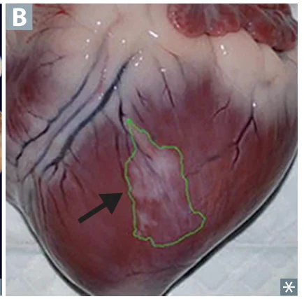
occurs in venous occlusion, in tissues with dual or collateral blood supply (liver, lung, intestine, testes), and with reperfusion (post-angioplasty). Blood re-enters loose necrotic tissue.Pale (anemic) infarct
occurs in solid organs with a single end-arterial supply (heart, kidney, spleen). There is no collateral to bleed in.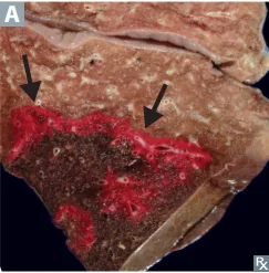
| Feature | Red Infarct | Pale Infarct |
|---|---|---|
| Blood supply | Dual/collateral, or venous occlusion | Single end-arterial |
| Organs | Lung, liver, intestine, testes | Heart, kidney, spleen |
| Setting | Venous occlusion, reperfusion | Arterial occlusion |
| Mechanism | Blood re-enters loose necrotic tissue | No collateral to bleed in |
Free radical injury
- Generated by radiation, phase I drug metabolism (P-450), redox reactions, NO, transition metals (iron and copper via the Fenton reaction), and the neutrophil/macrophage oxidative burst.
- Eliminated by scavenging enzymes (catalase, superoxide dismutase, glutathione peroxidase), antioxidants (vitamins A, C, E), spontaneous decay, and metal carrier proteins (transferrin, ceruloplasmin).
Classic free-radical diseases
Classic free-radical diseases
- O₂ toxicity in newborns: retinopathy of prematurity and bronchopulmonary dysplasia.
- Reperfusion injury after thrombolysis or stenting.
- Acetaminophen overdose, and carbon tetrachloride (P-450 → CCl₃• → fatty liver via ↓ apolipoprotein synthesis plus centrilobular necrosis).
- Metal overload: hemochromatosis (iron) and Wilson disease (copper).
After a prolonged episode of hypotension, a patient's liver enzymes rise sharply. Which part of the liver lobule is injured first, and why?
Zone III, around the central vein. It is furthest from the portal triad, so it receives the least oxygenated blood and is the first to die when perfusion falls.
How it's tested
After a severe hypotensive episode (shock, GI bleed, cardiac arrest), transaminases spike into the thousands. This is ischemic hepatic necrosis localized to Zone III (centrilobular), because zone III is furthest from the oxygenated blood of the portal triad.
Separately, the paradox question: cardiac enzymes rise further immediately after successful stenting or thrombolysis. This is reperfusion injury, where restored O₂ delivery fuels a free-radical burst, plus Ca²⁺ overload and neutrophil influx, that kills myocytes the original ischemia had merely stunned.
Go deeper
First Aid "Ischemia" + "Free radical injury"; Pathoma Ch. 1; Boards & Beyond. Anchor the ischemia-sensitive cells (hippocampal/Purkinje neurons, cardiac subendocardium, renal PCT straight segment + TAL) and free-radical mechanisms — lipid peroxidation, reperfusion injury, and the antioxidant scavengers.
High-yield images2
Flashcards for this page
Card 1 of 5 · try-out only, nothing is saved
(...) injury
You just read one page of 944
The rest of Step 1, written exactly like this.
Rapid Review is the reading layer of a full Step 1 platform: a schedule fitted to your exam date, flashcards for every page, and a mentor a message away.
Every Step 1 system
Written the same way: what to know, how it’s tested, where to go deeper.
A plan built to your exam date
Tell us when you sit, and the schedule fits the whole library and your question bank to it.
Flashcards for every page
Ready-made spaced-repetition decks linked to each topic, so nothing you read gets forgotten.
