
Rapid Review·General Pathology
Select any text to highlight it or add a note.
CELLULAR INJURY
T1Must knowNecrosis
P205
Focus on
Uncontrolled cell death that spills its contents and ignites inflammation, and matching the histologic pattern to the organ and the insult.
Key takeaways
What makes it necrosis
The defining sequence
exogenous injury → membrane damage → contents leak → enzymatic degradation plus protein denaturation → local inflammatory reaction. That inflammation is the key contrast with apoptosis.The six patterns
Coagulative: every infarct except the brain
What this shows
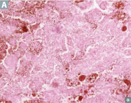
Coagulative
ischemia or infarct in any tissue except brain.- Injury denatures the lytic enzymes too, so proteolysis is blocked and cellular architecture is preserved as ghost outlines: nuclei disappear, cytoplasm is intensely eosinophilic.
- MI and acute tubular necrosis are the classics.
Liquefactive: brain and abscess
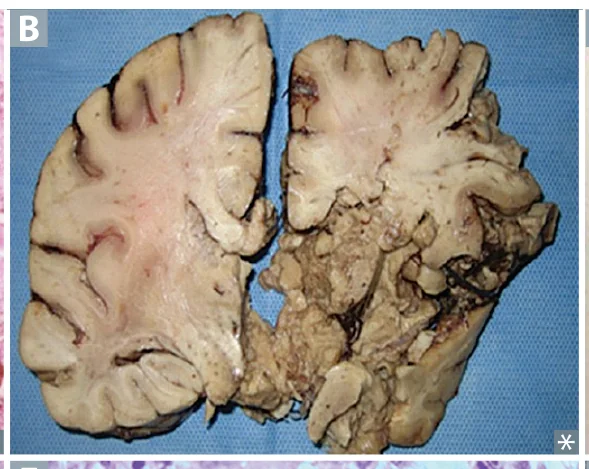
Liquefactive
CNS infarcts and bacterial abscesses.- Neutrophils release lysosomal enzymes that digest the tissue, so architecture is destroyed.
- Early: debris plus macrophages. Late: cystic cavitation.
Caseous: TB and fungi
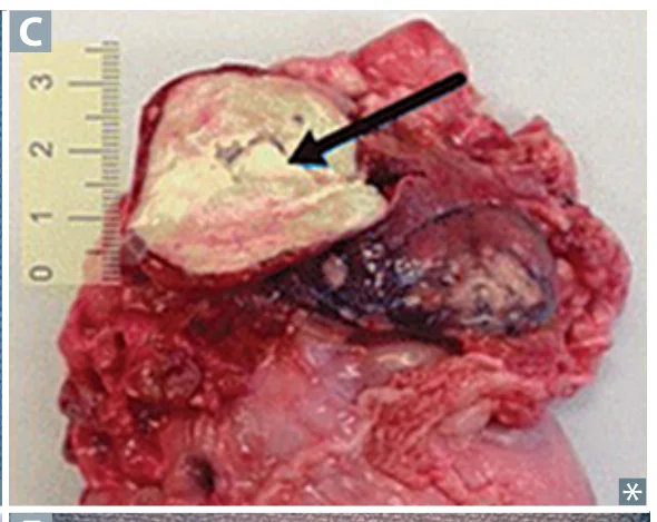
Caseous
TB, systemic fungi (Histoplasma), Nocardia, and Bartonella henselae (cat-scratch).- Macrophages wall off the organism → granular debris surrounded by lymphocytes and macrophages → cheese-like gross appearance.
Fat: pancreatitis and breast trauma
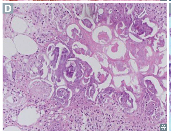
Fat, enzymatic
acute pancreatitis.- Lipase breaks down triglycerides → free fatty acids bind Ca²⁺ → saponification, chalky-white grossly and dark blue on H&E.
- Fat, nonenzymatic: breast trauma, no lipase involved.
Fibrinoid: vessel walls
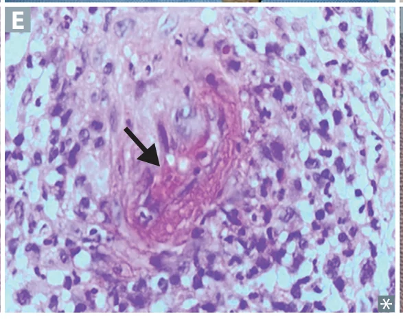
Fibrinoid
vessel walls contain an eosinophilic layer of proteinaceous material.- Immune (polyarteritis nodosa, type III hypersensitivity) or nonimmune (hypertensive emergency, preeclampsia).
- "-oid" means looks like fibrin but isn't.
Gangrenous: dry, wet and gas
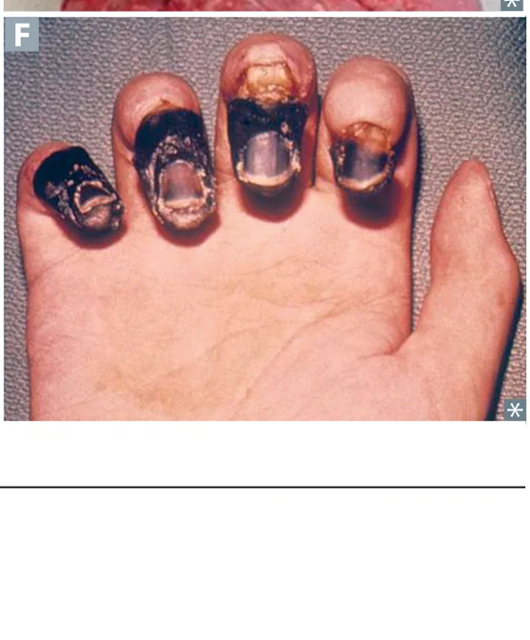
Gangrenous
distal extremity or GI tract after chronic ischemia.- Dry = coagulative (diabetic foot, Buerger disease in a young smoker).
- Wet = liquefactive superimposed on coagulative (superinfection; Fournier gangrene of the perineum in advanced diabetes).
- Gas gangrene = C. perfringens α-toxin/phospholipase → CO₂ in tissue → crepitus. Can also strike the gallbladder as emphysematous cholecystitis.
Cystic medial necrosis
Cystic medial necrosis
necrosis of large arteries → aortic dissection or aneurysm.- Seen in Marfan, Ehlers-Danlos, and chronic HTN. The NBME may write it simply as "medial necrosis," which is the same thing.
Side by side
| Necrosis Type | Mechanism | Histology / Gross | Classic Scenario |
|---|---|---|---|
| Coagulative | Enzymes denatured → proteolysis blocked | Architecture preserved, nuclei gone, ↑ eosinophilia | MI, ATN, pale infarcts |
| Liquefactive | Neutrophil lysosomal enzymes digest tissue | Cystic cavitation, pus | Brain infarct, abscess |
| Caseous | Macrophages wall off organism | Granuloma with central "cheese" | TB, Histoplasma, cat-scratch |
| Fat | Lipase → FFA plus Ca²⁺ saponification | Dead fat cells, chalky white / dark blue H&E | Pancreatitis; breast trauma |
| Fibrinoid | Immune complex plus fibrin leak | Pink proteinaceous vessel wall | PAN, malignant HTN, preeclampsia |
| Gangrenous | Chronic ischemia ± superinfection | Dry (coagulative) vs. wet (liquefactive) | Diabetic foot, Buerger |
During surgery for severe acute pancreatitis, chalky-white deposits are seen in the peripancreatic fat. What type of necrosis is this, and how do the deposits form?
Enzymatic fat necrosis. Pancreatic lipase breaks down triglycerides, and the released free fatty acids bind Ca²⁺ (saponification), giving chalky-white deposits grossly and a dark blue color on H&E.
How it's tested
The brain is the exception that carries the question. Every CNS infarct is liquefactive necrosis, never coagulative. Acutely, the buzzword histology is "red neurons" (intensely eosinophilic shrunken neurons within 12 to 24 hours), in direct contrast to the myocardium, where the answer immediately post-MI is "no histologic changes."
Microglia are the CNS macrophages that clear the debris, and astrocytes are the cells that form the glial scar (gliosis). There are no fibroblasts making collagen in the brain, so "fibroblast" is always wrong for CNS scarring.
Go deeper
First Aid "Necrosis" table; Pathoma Ch. 1 "Necrosis"; Boards & Beyond. Anchor each type to its trigger organ — coagulative (heart/kidney/most organs), liquefactive (brain + abscess), caseous (TB/fungi), fat (pancreas/breast), fibrinoid (vessels, type III), gangrene (limbs/gut).
High-yield images6
Flashcards for this page
Card 1 of 5 · try-out only, nothing is saved
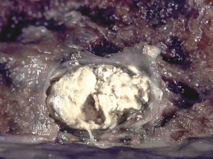
Fragmented cells and debris surrounded by (...) and (...)
You just read one page of 944
The rest of Step 1, written exactly like this.
Rapid Review is the reading layer of a full Step 1 platform: a schedule fitted to your exam date, flashcards for every page, and a mentor a message away.
Every Step 1 system
Written the same way: what to know, how it’s tested, where to go deeper.
A plan built to your exam date
Tell us when you sit, and the schedule fits the whole library and your question bank to it.
Flashcards for every page
Ready-made spaced-repetition decks linked to each topic, so nothing you read gets forgotten.
