
Rapid Review·Cardiovascular
Select any text to highlight it or add a note.
Vascular: Aorta & Carotid
T1Must knowAortic Dissection
Focus on
Sudden tearing chest pain radiating to the back in a hypertensive man — or a young Marfan patient — with unequal pulses. Chest film, then CT angiography (TEE if unstable). IV beta blocker first to a heart rate under 60 and SBP 100–120, then nitroprusside, never the vasodilator alone. Type A goes to surgery; type B is managed medically unless complicated.
Key takeaways
What it is and who gets it
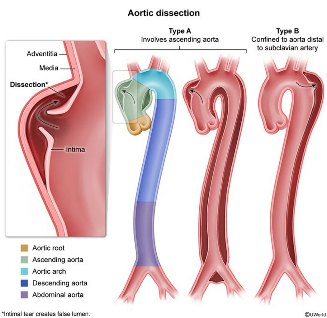
What this shows
Aortic dissection
a tear in the intima lets pulsatile blood separate the layers of the aortic wall, creating a false lumen that propagates and compresses the true lumen and any branch it reaches.Stanford A
involves the ascending aorta (with or without the descending); a surgical emergency.Stanford B
involves the descending aorta distal to the left subclavian only; medical unless complicated.Hypertension is the strongest risk factor
and the most common cause over 60; also thoracic aortic aneurysm and atherosclerosis.Other risk factors
connective-tissue disorders (Marfan, the common cause under 40; Ehlers-Danlos), bicuspid aortic valve, Turner syndrome, syphilitic aortitis, cocaine and amphetamines, trauma.Clinical features
The pain
severe anterior chest pain, tearing or ripping, radiating to the interscapular back; syncope, diaphoresis, confusion or agitation.Asymmetry
a pressure difference over 20 mm Hg between the arms, a wide pulse pressure, a pulse deficit.Blood pressure
hypertension is usual; hypotension means rupture, tamponade or acute aortic regurgitation.Extension findings
a new diastolic murmur (aortic regurgitation), ST elevation from a coronary ostium (usually the right), stroke, Horner syndrome, hoarseness, limb, renal or mesenteric ischemia.Diagnosis
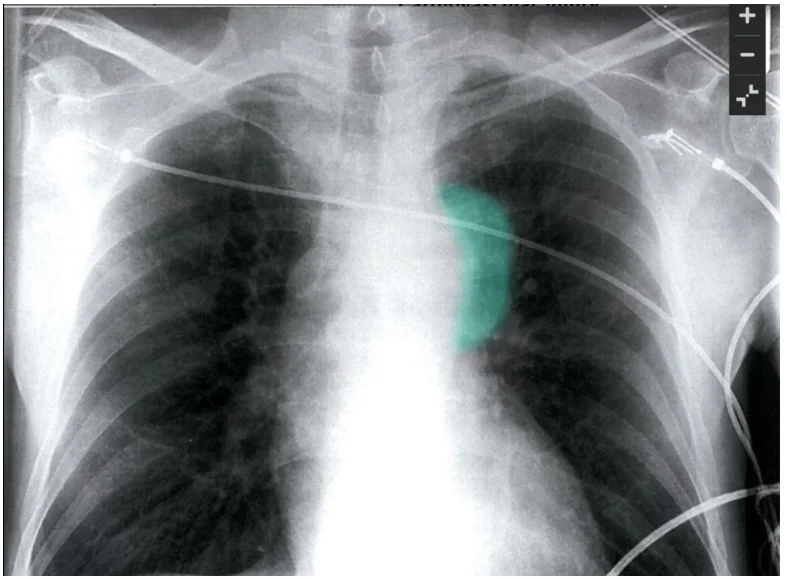
Electrocardiogram and chest radiograph for every acute chest pain
the film shows a widened mediastinum and an abnormal aortic knob, but is normal in 10 to 20 percent.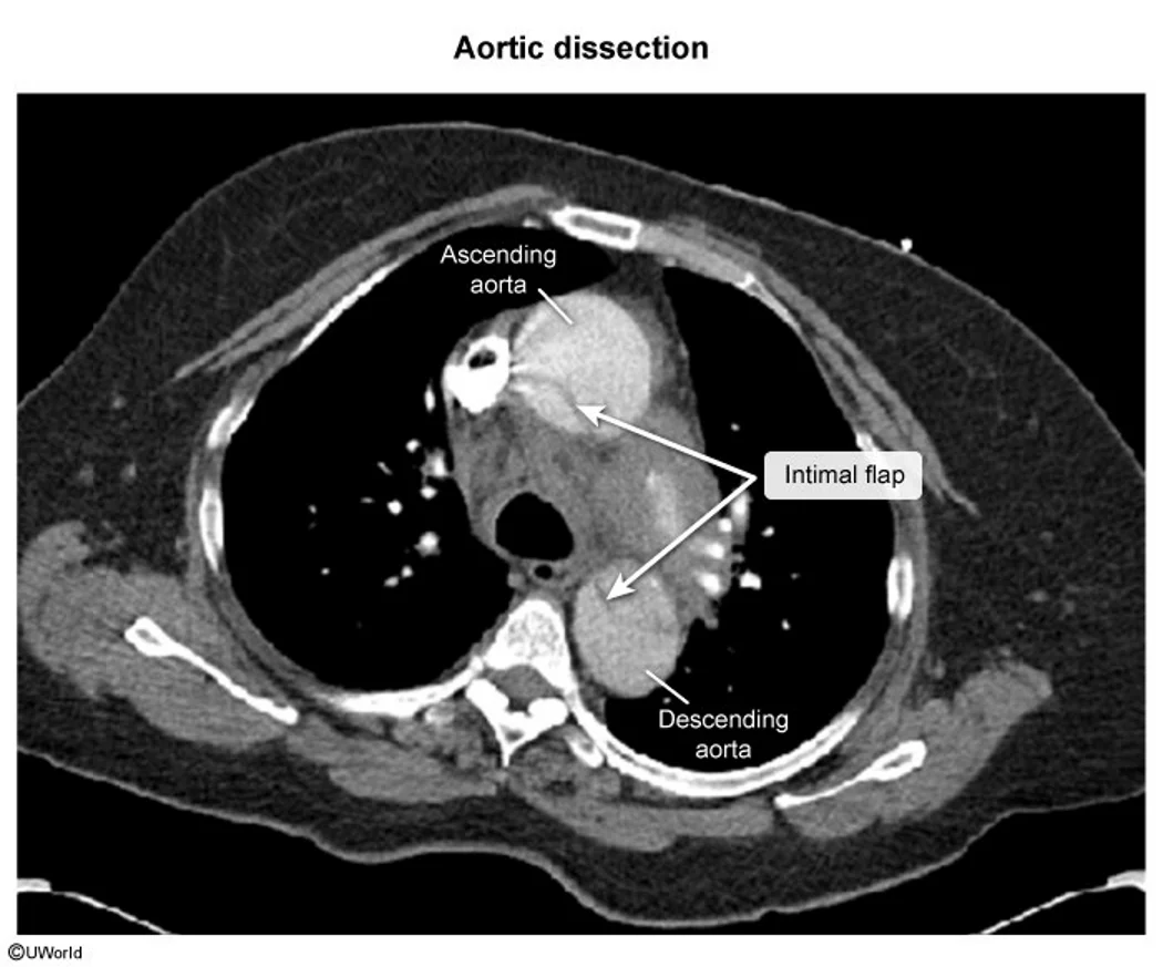
What this shows
Confirm by stability
per the Follow the diagnostic pathway.- Computed tomography (CT) angiography in the stable patient (check creatinine and contrast allergy first).
- Transesophageal echocardiography (TEE) in the unstable patient or when contrast is contraindicated (kidney disease, allergy).
| Type A | Type B | |
|---|---|---|
| Location | Ascending aorta, with or without the descending | Descending aorta beyond the left subclavian |
| Pain | Anterior chest, neck, jaw | Interscapular back, abdomen |
| Complications (by extension) | Aortic regurgitation, coronary occlusion (infarction), cardiac tamponade, stroke, Horner syndrome, vocal cord paralysis | Limb, renal and mesenteric ischemia |
| Treatment | Anti-impulse therapy plus immediate surgery (open or endovascular repair) | Medical anti-impulse therapy; endovascular repair (TEVAR) if complicated or progressing |
Anti-impulse therapy for everyone
Goals
pain control, and a lower heart rate and systolic pressure to cut left-ventricular contractility and aortic wall stress.Targets
heart rate under 60 to 80 and systolic 100 to 120 mm Hg, as in the Use the Type A versus Type B management fork.Intravenous beta blocker first
esmolol, labetalol; a non-dihydropyridine calcium-channel blocker if beta blockers are contraindicated.Add nitroprusside
only if the systolic pressure stays over 120 despite beta blockade.Analgesia
intravenous opioids (pain drives the catecholamines).Hypotension
fluids and blood, look for tamponade or rupture; pericardiocentesis is avoided (it can accelerate bleeding), so go to the operating room.Definitive treatment
Type A: emergency surgery
ascending aortic graft, with valve or root replacement when involved.Type B: medical therapy
with thoracic endovascular aortic repair (TEVAR) for complications (malperfusion, rupture, refractory pain or hypertension, rapid expansion).Long-term
tight blood pressure control (beta blocker), serial imaging for aneurysmal degeneration of the false lumen, genetic evaluation in the young or syndromic.A 58-year-old hypertensive man has sudden tearing chest pain radiating between the shoulder blades and a 30 mm Hg difference between the arms; he is stable and his creatinine is normal. What is the confirmatory test, the first drug, and the treatment if the ascending aorta is involved?
Aortic dissection: CT angiography (TEE if unstable). Start an intravenous beta blocker (esmolol, labetalol) to a heart rate under 60 to 80 and systolic 100 to 120, adding nitroprusside only after beta blockade. Ascending involvement (Stanford A) means emergency surgery.
How it's tested
A 65-year-old hypertensive man with sudden tearing chest pain radiating between the shoulder blades, BP 190/100 right arm and 150/90 left, widened mediastinum: aortic dissection — CT angiography; IV esmolol now, nitroprusside once the rate is controlled.
Same story with BP 80/50, JVD and muffled heart sounds: type A rupturing into the pericardium — TEE and the operating room; not routine pericardiocentesis, not heparin.
Tearing pain with inferior ST elevation: dissection involving the right coronary ostium — do not give thrombolytics or heparin; CT angiography or TEE first.
Dissection confined to the descending aorta, pain controlled, no malperfusion: type B — medical management with beta blockade; TEVAR only if complicated.
Which drug is given first and why: a beta blocker — lowering pressure with a vasodilator alone causes reflex tachycardia that increases shear stress and propagates the tear.
A 28-year-old tall man with pectus excavatum and chest pain: Marfan syndrome — dissection until proven otherwise; also image the root for aneurysm.
Go deeper
Guidelines: 2022 ACC/AHA Aortic Disease Guideline
Related Step 2 pages: Approach to Aortic Syndromes, Thoracic Aortic Aneurysm, Aortic Regurgitation, Cardiac Tamponade, Hypertension, Approach to Chest Pain, Bicuspid Aortic Valve
High-yield images5
Flashcards for this page
Card 1 of 5 · try-out only, nothing is saved
You just read one page of 236
The rest of Step 2 CK, written exactly like this.
Rapid Review is the reading layer of a full Step 1 platform: a schedule fitted to your exam date, flashcards for every page, and a mentor a message away.
Every Step 1 system
Written the same way: what to know, how it’s tested, where to go deeper.
A plan built to your exam date
Tell us when you sit, and the schedule fits the whole library and your question bank to it.
Flashcards for every page
Ready-made spaced-repetition decks linked to each topic, so nothing you read gets forgotten.
