
Rapid Review·Cardiovascular
Select any text to highlight it or add a note.
Vascular: Aorta & Carotid
T1Must knowAbdominal Aortic Aneurysm
Focus on
A male smoker over 65 with a pulsatile abdominal mass. Screen once with ultrasound, watch it until 5.5 cm or growth of more than 0.5 cm in 6 months, then repair. Rupture is abdominal or back pain, shock and a pulsatile mass: known aneurysm and unstable — the operating room; unknown and unstable — bedside ultrasound; stable — CT angiography.
Key takeaways
What it is and who gets it
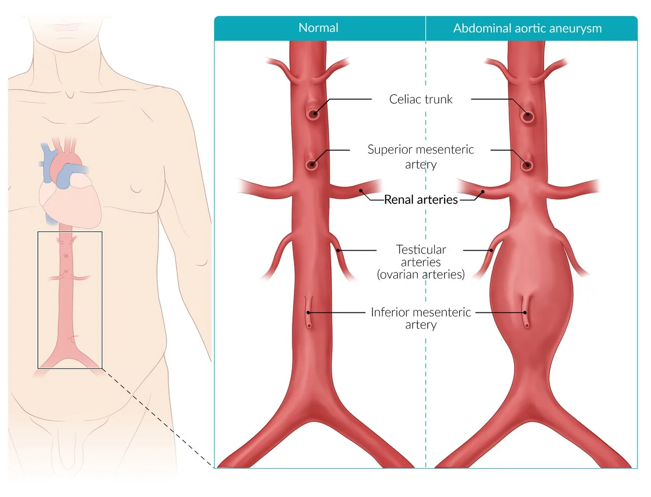
What this shows
Abdominal aortic aneurysm (AAA)
a focal dilation of all three layers of the abdominal aorta to 3 cm or more, most often infrarenal and fusiform.Laplace
wall tension rises with radius, so the larger the aneurysm the faster it grows and the more likely it ruptures.Smoking is the number one risk factor
for development, growth and rupture.Other risk factors
age over 60, male sex, hypertension, hyperlipidemia, atherosclerotic disease, family history.Diabetes is protective
lower incidence and growth, for unclear reasons.Clinical features, screening and diagnosis
Asymptomatic when stable
found on imaging done for something else or on screening.Symptomatic but not ruptured
back pressure or pain, a pulsatile abdominal mass (present in just over half), prevertebral calcification on a plain film.Screening
a one-time ultrasound for men 65 to 75 who have ever smoked (United States Preventive Services Task Force (USPSTF)).Abdominal ultrasound
for asymptomatic patients (screening) and for symptomatic unstable patients (bedside, fast, no contrast).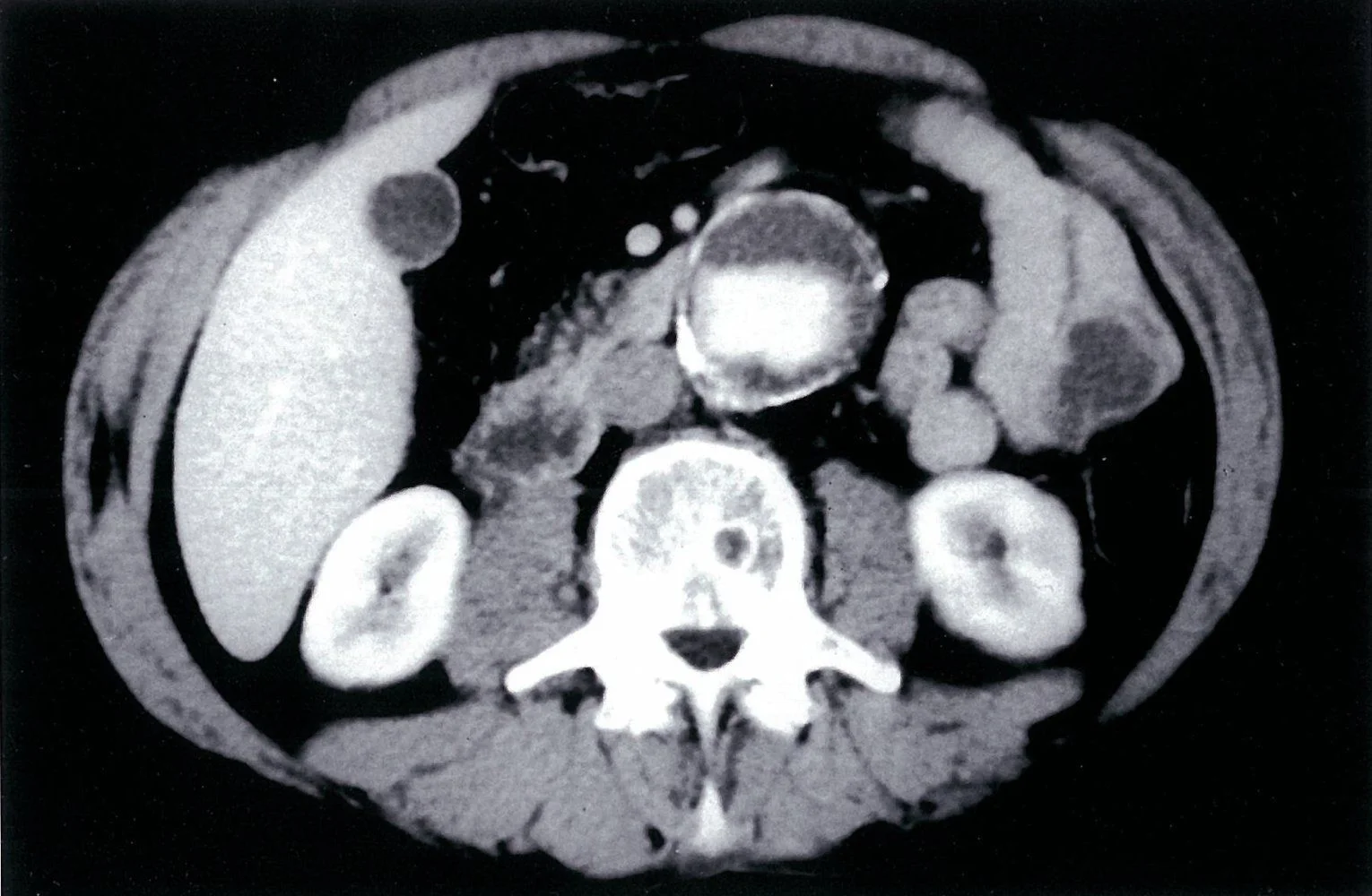
What this shows
Computed tomography (CT) angiography
for symptomatic stable patients and for operative planning; the Recognize ruptured AAA on CT shows the leak.| Situation | Test |
|---|---|
| Screening or surveillance, asymptomatic | Abdominal ultrasound |
| Symptomatic and stable | CT angiography |
| Unstable, no known aneurysm | Bedside ultrasound, then the operating room if positive |
| Unstable with a known aneurysm | Operating room directly, no imaging (time is survival) |
Management of the unruptured aneurysm
Risk-factor modification for all
smoking cessation (the best intervention against progression) and statins.Asymptomatic under 5.5 cm
surveillance ultrasound every 6 to 12 months.Repair (endovascular or open)
for a diameter of 5.5 cm or more, rapid expansion (over 1 cm/year or over 0.5 cm in 6 months), or any symptoms (elective repair).Rupture
Where it ruptures
usually posterior into the retroperitoneum, which contains the bleed and can delay instability by an hour or more; anterior rupture into the peritoneum causes rapid shock.The classic triad
abdominal pain, shock and a pulsatile abdominal mass.Strongest predictors of rupture
current smoking, diameter over 5.5 cm, expansion over 1 cm/year, hypertension and female sex.Imaging by stability
stable with a known aneurysm, confirm with CT angiography; unstable without a known aneurysm, bedside ultrasound, as on the Use the unstable-AAA pathway.Treatment
emergent endovascular or open repair, with permissive hypotension and blood products until the aorta is controlled.Complications of repair
Ischemic colitis
from inferior mesenteric artery ligation: progressive abdominal pain and bloody diarrhea in the first days; sigmoidoscopy.Aortoenteric fistula
significant gastrointestinal bleeding years after a graft (a herald bleed, then massive); CT angiography and urgent surgery.Others
endoleak after endovascular repair (lifelong imaging), graft infection, myocardial infarction (the leading peri-operative death).A 70-year-old former smoker has an asymptomatic 4.8 cm infrarenal aneurysm that measured 4.0 cm a year ago. What is the management, and what is the single best step against progression?
Repair (endovascular or open): it has expanded over 1 cm in a year, even though it is under 5.5 cm. The best intervention against progression is smoking cessation, plus a statin.
How it's tested
A 67-year-old man who quit smoking ten years ago asks about screening: a one-time abdominal ultrasound — he "ever smoked".
A 4.5-cm AAA found incidentally: stop smoking, statin, blood pressure control, and ultrasound every 12 months; repair at 5.5 cm or if it grows > 0.5 cm in 6 months.
Severe back pain, BP 80/40, a pulsatile epigastric mass, no known aneurysm: bedside ultrasound, then emergency repair — not a CT.
Same presentation in a man with a known 6-cm aneurysm: straight to the operating room.
Bloody diarrhea and abdominal pain on day 2 after open AAA repair: ischemic colitis from IMA ligation — sigmoidoscopy.
Massive hematemesis three years after an aortic graft: aortoenteric fistula — CT angiography, urgent surgery.
Go deeper
Guidelines: 2022 ACC/AHA Aortic Disease Guideline · USPSTF AAA Screening Recommendation
Related Step 2 pages: Approach to Aortic Syndromes, Thoracic Aortic Aneurysm, Aortic Dissection, Hypovolemic & Hemorrhagic Shock, Peripheral Artery Disease, Approach to Preoperative Cardiac Evaluation
High-yield images4
Flashcards for this page
Card 1 of 5 · try-out only, nothing is saved
You just read one page of 236
The rest of Step 2 CK, written exactly like this.
Rapid Review is the reading layer of a full Step 1 platform: a schedule fitted to your exam date, flashcards for every page, and a mentor a message away.
Every Step 1 system
Written the same way: what to know, how it’s tested, where to go deeper.
A plan built to your exam date
Tell us when you sit, and the schedule fits the whole library and your question bank to it.
Flashcards for every page
Ready-made spaced-repetition decks linked to each topic, so nothing you read gets forgotten.
