
Rapid Review·Cardiovascular
Select any text to highlight it or add a note.
Congenital Heart Disease
T1Must knowAtrial Septal Defect
Focus on
A soft ejection murmur at the left upper sternal border with a widely split S2 that does not move with breathing — the fixed split is the diagnosis. A low-pressure shunt that stays silent for decades, then declares itself as atrial fibrillation, right heart failure, pulmonary hypertension or a paradoxical embolic stroke. Close it when the RV is enlarged, before pulmonary vascular disease makes closure impossible.
Key takeaways
What it is and who gets it
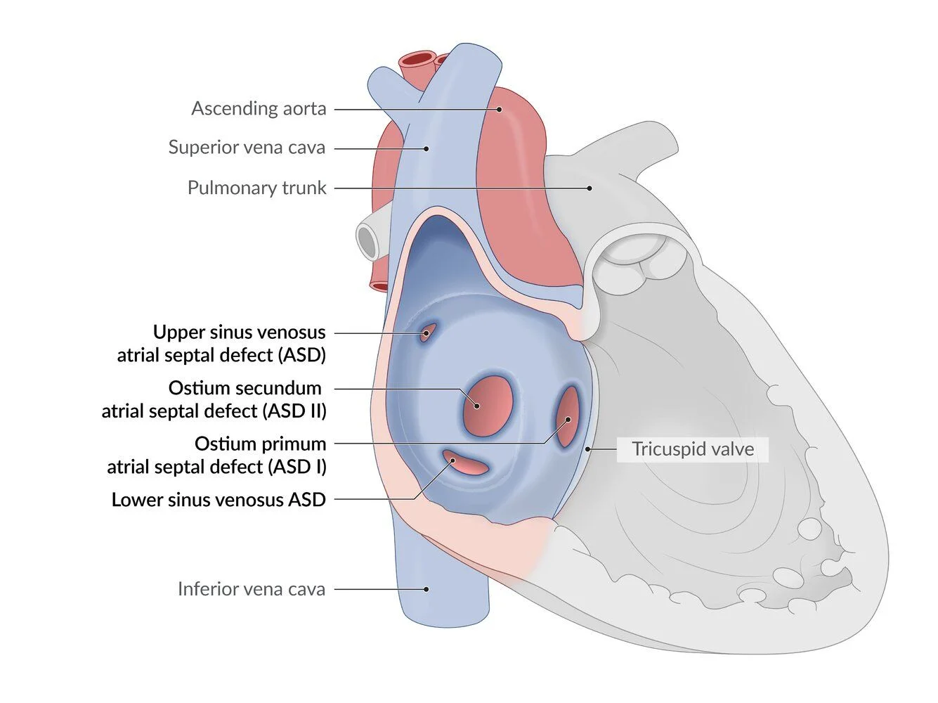
What this shows
Atrial septal defect
a tissue deficiency in the atrial septum allowing a continuous low-pressure, low-volume left-to-right shunt, so the right heart and lungs are volume-loaded slowly.Ostium secundum (much more common)
arrested growth of the septum secundum or excessive absorption of the septum primum.Ostium primum
the septum primum fails to fuse with the endocardial cushions; part of the atrioventricular canal spectrum with a cleft mitral valve.Not an atrial septal defect
a Patent Foramen Ovale.Risk factors
Down syndrome (primum defects), fetal alcohol syndrome, intrauterine infections.Clinical features and diagnosis
Usually asymptomatic until middle age
when pulmonary hypertension, atrial arrhythmias or Eisenmenger physiology appear.- Only large defects cause heart failure symptoms in childhood (dyspnea, fatigue, exercise intolerance).
Examination
four findings.- A systolic ejection murmur at the pulmonary area (functional pulmonic stenosis from the extra flow; the shunt itself is silent).
- Wide fixed splitting of S2.
- A mid-diastolic rumble at the tricuspid area with large shunts.
- A right-ventricular heave.
Paradoxical embolism
a venous thrombus crosses the defect into the arterial circulation and causes an ischemic stroke.Transthoracic echocardiography is confirmatory
type, size, shunt direction, right heart enlargement, pulmonary pressure; transesophageal echo before device closure.Medium to large defects
right ventricular hypertrophy on the electrocardiogram, increased pulmonary vascular markings on the film.- Secundum: right axis and incomplete right bundle branch block; primum: left axis with first-degree block.
| Ostium secundum | Ostium primum | Patent Foramen Ovale | |
|---|---|---|---|
| Site | Mid-septum, fossa ovalis | Low septum, next to the atrioventricular valves | Fossa ovalis; a flap, not a hole |
| Association | Sporadic, fetal alcohol | Down syndrome, atrioventricular canal, cleft mitral valve | About 25 percent of the population |
| Electrocardiogram | Right axis, incomplete right bundle branch block | Left axis, first-degree block | Normal |
| Closure | Transcatheter device | Surgical patch with mitral repair | Only after an embolic event |
Management and complications
Children, small and asymptomatic
serial echocardiography; up to 40 percent close spontaneously by age 5.Large or symptomatic
closure, transcatheter for secundum defects with adequate rims, surgical patch for primum and sinus venosus defects.Adults
close for symptoms, right-ventricular overload, or an embolic stroke.Complications
atrial fibrillation and flutter in adults, paradoxical embolic stroke, pulmonary hypertension and Eisenmenger syndrome, right heart failure, mitral regurgitation with primum defects.A 35-year-old woman has new palpitations; she has a soft systolic ejection murmur at the left upper sternal border and a second heart sound that stays widely split in both inspiration and expiration. What is the lesion, the confirmatory test, and when is closure contraindicated?
An ostium secundum atrial septal defect (flow murmur plus wide fixed split S2; atrial arrhythmias appear in adulthood). Transthoracic echocardiography confirms it; closure is contraindicated once severe pulmonary arterial hypertension has developed.
How it's tested
A 7-year-old with a soft ejection murmur at the left upper sternal border and a widely split S2 that does not change with respiration; ECG shows right axis deviation and rSR′ in V1: secundum ASD — echocardiogram; device closure if the RV is enlarged.
A 38-year-old woman with new atrial fibrillation, exertional dyspnea and a fixed split S2: an undiagnosed ASD — echo; close it after rate control and anticoagulation if pulmonary resistance allows.
A young adult with a cryptogenic stroke and a DVT; echo shows an atrial-level shunt: paradoxical embolism through an ASD (or PFO) — closure.
Child with Down syndrome, fixed split S2, left axis deviation and an apical holosystolic murmur: primum ASD with a cleft mitral valve — surgical repair.
Why must closure be avoided in Eisenmenger physiology: the defect is the RV's escape route; closing it against fixed pulmonary hypertension causes right heart failure.
Go deeper
Guidelines: 2018 AHA/ACC Adult Congenital Heart Disease Guideline
Related Step 2 pages: Patent Foramen Ovale, Ventricular Septal Defect, Eisenmenger Syndrome, Heart Sounds & Murmurs, Cardiac Embryology Essentials, Atrial Fibrillation
High-yield images1
You just read one page of 236
The rest of Step 2 CK, written exactly like this.
Rapid Review is the reading layer of a full Step 1 platform: a schedule fitted to your exam date, flashcards for every page, and a mentor a message away.
Every Step 1 system
Written the same way: what to know, how it’s tested, where to go deeper.
A plan built to your exam date
Tell us when you sit, and the schedule fits the whole library and your question bank to it.
Flashcards for every page
Ready-made spaced-repetition decks linked to each topic, so nothing you read gets forgotten.
