
Rapid Review·Cardiovascular
Select any text to highlight it or add a note.
Foundations
T1Must knowCardiac Anatomy
Videos
1 to play here
Play 1 here
Focus on
Step 2 tests anatomy only through its consequences: which coronary feeds which wall (and which leads light up), which chamber sits where on a chest film, where each valve is heard, and why one papillary muscle ruptures after an inferior MI.
Key takeaways
Where the heart sits
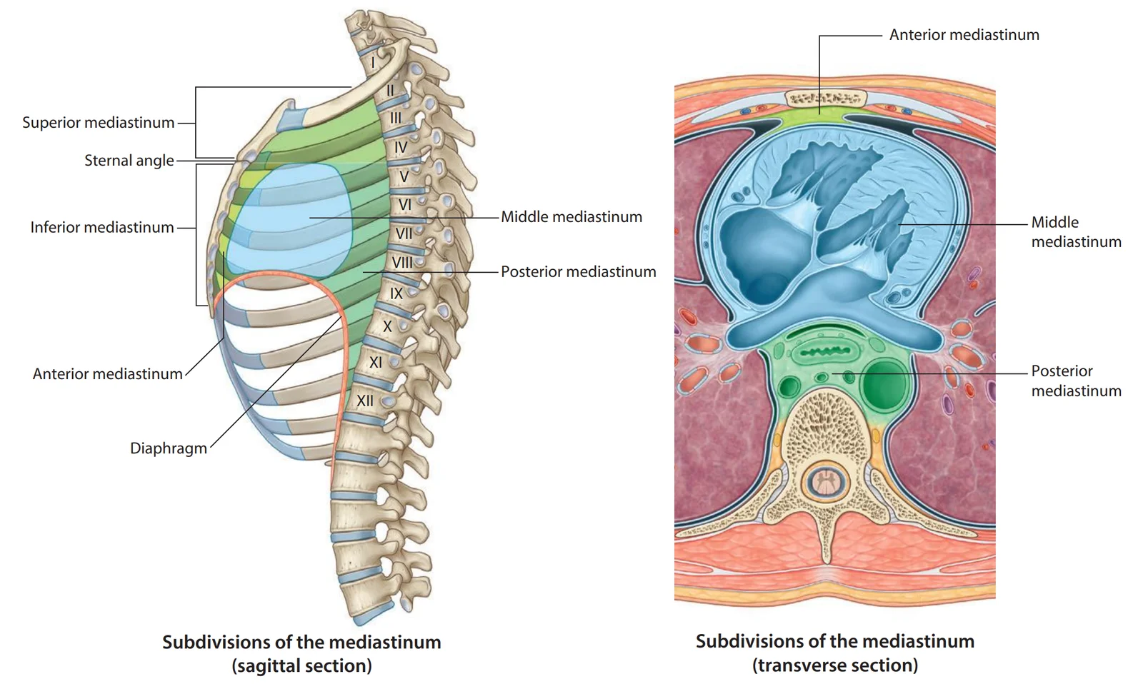
What this shows
Middle mediastinum
the heart sits in the middle mediastinum, inside the pericardial sac, between the two pleural cavities.- The anterior mediastinum (thymus, fat, nodes) borders it in front.
- The posterior mediastinum (esophagus, descending aorta, azygos, thoracic duct) borders it behind.
Pericardium, from outside in
- Fibrous pericardium: the tough outer sac.
- Parietal serous pericardium: lines the inside of the fibrous sac.
- Pericardial space: the thin gap between the two serous layers.
- Visceral pericardium (epicardium): stuck to the heart surface.
Parietal layer is phrenic-innervated
which is why pericarditis pain refers to the neck and shoulder.Right ventricle (RV) is the most anterior chamber
it sits directly behind the sternum, as the anterior surface of the heart shows.- So it is the chamber most often injured in blunt chest trauma (Blunt Cardiac Injury).
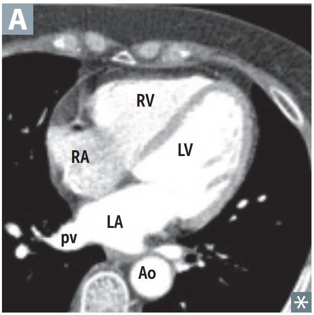
What this shows
Left atrium (LA) is the most posterior chamber
it lies against the esophagus.- LA enlargement (classically Mitral Stenosis) presses on the esophagus and causes dysphagia.
- It can also stretch the left recurrent laryngeal nerve and cause hoarseness (Ortner syndrome).
- It is also why a transesophageal echo (TEE) sees the LA and mitral valve best.
Inferior (diaphragmatic) surface
roughly two-thirds left ventricle (LV) and one-third RV.| Chamber | Position | Clinical consequence |
|---|---|---|
| Left atrium | Most posterior | Dysphagia and hoarseness when enlarged; best seen on TEE |
| Right ventricle | Most anterior | Most often injured in blunt trauma; forms the inferior border on a frontal film |
| Left ventricle | Left and inferior | Forms the left heart border; apex beat displaced when dilated |
Valves and where to listen
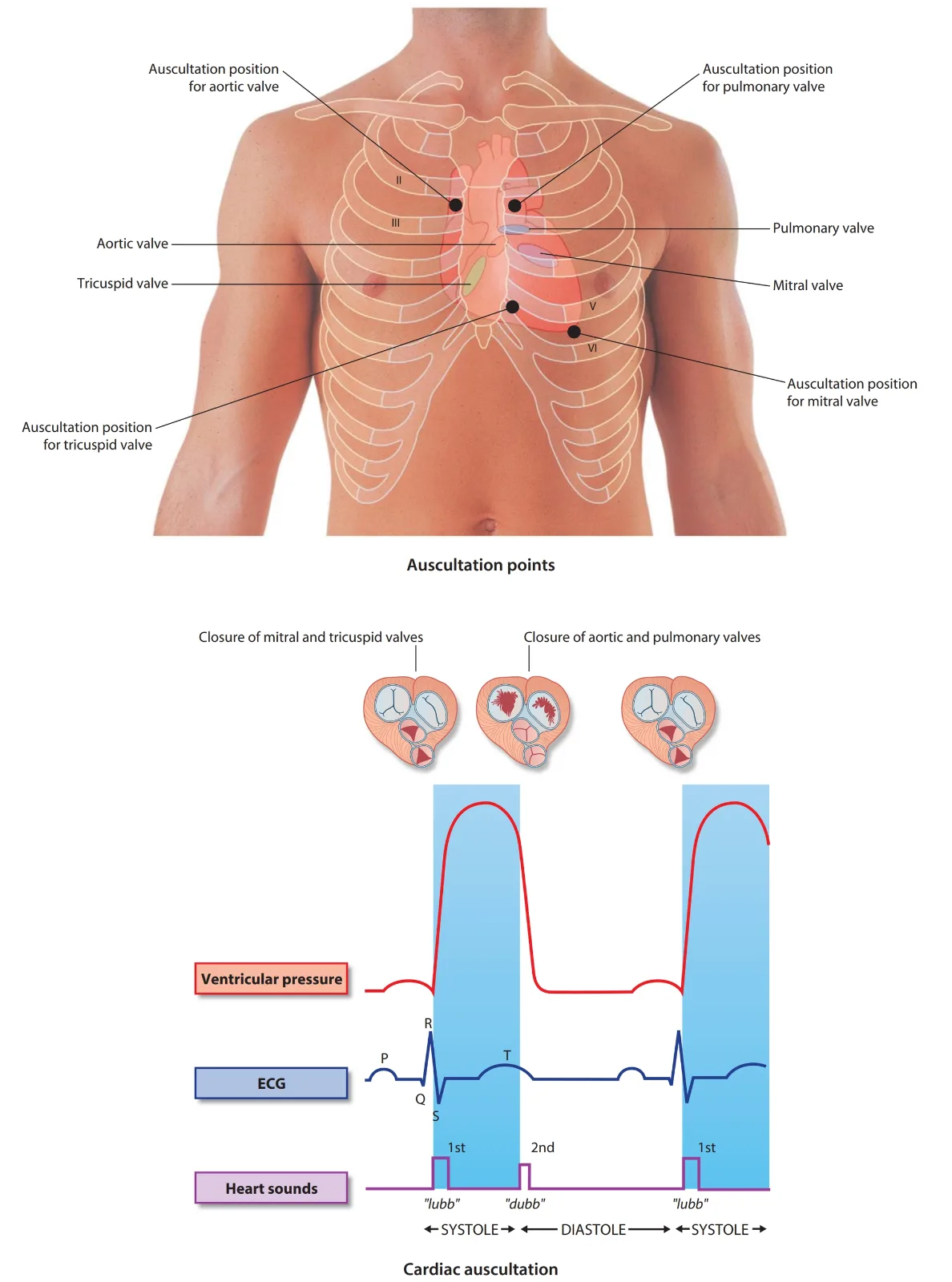
What this shows
Auscultation sites lie downstream of each valve
in the direction of flow, not directly over it.| Valve | Where heard | Association to remember |
|---|---|---|
| Aortic | 2nd right intercostal space, parasternal | Bicuspid valve (Turner syndrome); age-related sclerosis/calcific stenosis |
| Pulmonary | 2nd left intercostal space, parasternal | Stenosis is almost always congenital; rarely carcinoid heart disease |
| Tricuspid | 4th left intercostal space, parasternal | Most common valve in endocarditis of people who inject drugs |
| Mitral | 5th left intercostal space, midclavicular line (apex) | Most common valve in endocarditis overall; mitral stenosis is the classic rheumatic lesion |
Coronary arteries and what each occlusion does
Compare the two views
the anterior coronary view with the posterior (left-dominant) view.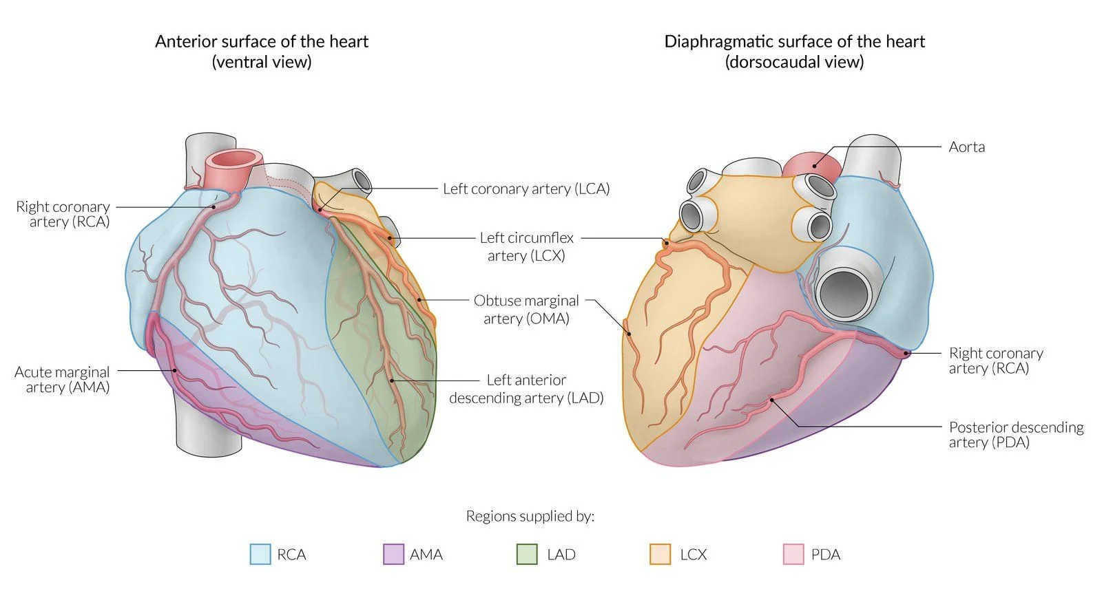
What this shows
Left anterior descending artery (LAD)
supplies the anterior two-thirds of the septum, the anterior LV wall and the anterolateral papillary muscle.- It is the most commonly occluded artery.
- Occlusion gives an anterior myocardial infarction (MI) (V1 to V4).
- This is the territory that most often produces pump failure and cardiogenic shock.
Left circumflex artery (LCX)
supplies the lateral and posterior LV (I, aVL, V5 to V6).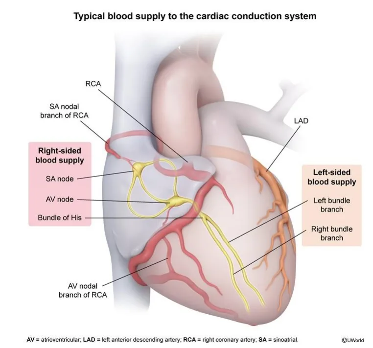
What this shows
Right coronary artery (RCA)
supplies the sinoatrial (SA) node (~60%) and the atrioventricular (AV) node (~90%) through its nodal branches.- It supplies the RV through the right (acute) marginal artery.
- So an inferior MI (II, III, aVF) brings sinus bradycardia and AV block, and can extend to the RV.
Posterior descending artery (PDA)
supplies the posterior third of the septum, the posterior two-thirds of the ventricular walls and the posteromedial papillary muscle.- The posteromedial muscle has this single supply.
- So it is the one that ruptures 2 to 7 days after an inferior MI, producing acute mitral regurgitation (Post MI Complications).
Dominance is defined by which artery gives off the PDA
right-dominant (RCA, ~85%, most common), left-dominant (LCX), codominant (both).| Pattern | PDA arises from | Frequency |
|---|---|---|
| Right-dominant | RCA | ~85%, most common |
| Left-dominant | LCX | ~5 to 10% |
| Codominant | Both RCA and LCX | ~10 to 20% |
| Papillary muscle | Blood supply | Rupture risk |
|---|---|---|
| Posteromedial | Single, PDA only | High, the classic rupture after an inferior MI |
| Anterolateral | Dual, LAD and LCX | Low |
Timing of flow
LV and septal perfusion happens mainly in early diastole; the RV is perfused throughout the cycle.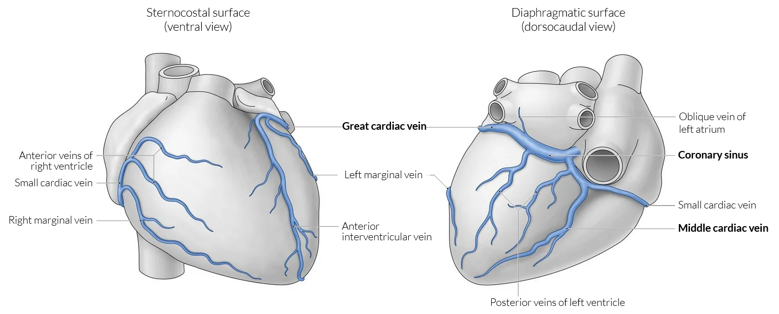
What this shows
Coronary sinus
the largest cardiac vein; it runs in the posterior left AV groove and drains into the right atrium (RA).- A dilated coronary sinus on echo suggests raised right-sided pressures (pulmonary hypertension) or a persistent left superior vena cava (SVC).
Right atrium and the conduction system
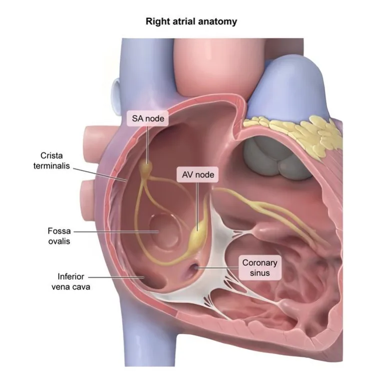
What this shows
Right atrial landmarks
seen on the opened right atrium.- The SA node sits at the crista terminalis near the SVC.
- The AV node sits in the interatrial septum near the coronary sinus ostium.
- The fossa ovalis is the closed foramen ovale.
- The coronary sinus opens beside the inferior vena cava (IVC).
Conduction runs in one direction
as drawn on the cardiac conduction system.SA node
fires first.Atria
the impulse spreads across both atria.AV node
delays it.Bundle of His
carries it into the septum.Right and left bundle branches
split it to each ventricle.Purkinje network
spreads it through the ventricular muscle.Details elsewhere
rates, intervals and electrocardiogram (ECG) interpretation live on ECG Basics.Reading the cardiac silhouette
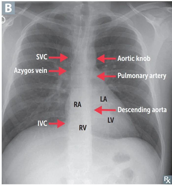
What this shows
Frontal film
each border is one structure.- Right border: right atrium.
- Left border: left ventricle, with the left atrial appendage above it.
- Inferior border: right ventricle.
- Superior border: atria and great vessels.
- Aortic knob: the aortic arch seen end-on; widening suggests aneurysm or dissection.
Lateral film
the front and back of the heart.- Anterior border: right ventricle (behind the sternum).
- Posterior border: left atrium above and left ventricle below.
Pattern shapes worth a glance
each classic silhouette points to one diagnosis.- LA enlargement splays the carina and gives a "double density".
- Boot-shaped heart: Tetralogy of Fallot.
- Egg-on-a-string: Transposition of the Great Arteries.
- Snowman: Total Anomalous Pulmonary Venous Return.
- Water-bottle heart: large Pericardial Effusion.
An inferior MI causes a heart rate of 40 on day 1 and a new holosystolic murmur with pulmonary edema on day 5. Which artery explains both, and why?
The right coronary artery. It supplies the AV node (~90%) and the SA node (~60%), so its occlusion brings bradycardia and AV block. In the usual right-dominant heart (~85%) it also gives off the posterior descending artery, the only supply of the posteromedial papillary muscle, which ruptures 2 to 7 days later and causes acute mitral regurgitation.
How it's tested
Chest pain with ST elevation in II, III and aVF, then a heart rate of 40 or a dropped beat: the RCA feeds the SA and AV nodes, so an inferior MI brings bradyarrhythmia; treat symptomatic bradycardia with atropine and check right-sided leads for RV involvement.
A new holosystolic murmur and pulmonary edema five days after an inferior MI: rupture of the posteromedial papillary muscle, the one with a single PDA supply — urgent echo and surgery.
A woman with mitral stenosis develops dysphagia and a hoarse voice: the enlarged left atrium, the most posterior chamber, is compressing the esophagus and the left recurrent laryngeal nerve.
A steering-wheel injury with new arrhythmia and raised troponin: the right ventricle sits directly behind the sternum and is the chamber most often contused.
Go deeper
Related Step 2 pages: ECG Basics, Acute Coronary Syndrome, Post MI Complications, Heart Sounds & Murmurs, Blunt Cardiac Injury, Fetal Circulation & Transition at Birth
High-yield images12
You just read one page of 236
The rest of Step 2 CK, written exactly like this.
Rapid Review is the reading layer of a full Step 1 platform: a schedule fitted to your exam date, flashcards for every page, and a mentor a message away.
Every Step 1 system
Written the same way: what to know, how it’s tested, where to go deeper.
A plan built to your exam date
Tell us when you sit, and the schedule fits the whole library and your question bank to it.
Flashcards for every page
Ready-made spaced-repetition decks linked to each topic, so nothing you read gets forgotten.
