
Rapid Review·Cardiovascular
Select any text to highlight it or add a note.
Foundations
T3Take a glanceCardiac Embryology Essentials
Videos
4 to play here
Play 4 here
Focus on
For CK, embryology matters only where it explains a congenital lesion: neural crest failure gives the conotruncal defects, septation failure gives ASD/VSD/AV canal, and the derivative table tells you what a persistent structure is.
Key takeaways
Looping and the outflow tract
Looping sets left-right polarity
the primary heart tube loops in week 4.- A defect in left-right dynein causes dextrocardia.
- With situs inversus it is Kartagener syndrome (primary ciliary dyskinesia): recurrent sinusitis, bronchiectasis, infertility.
What this shows
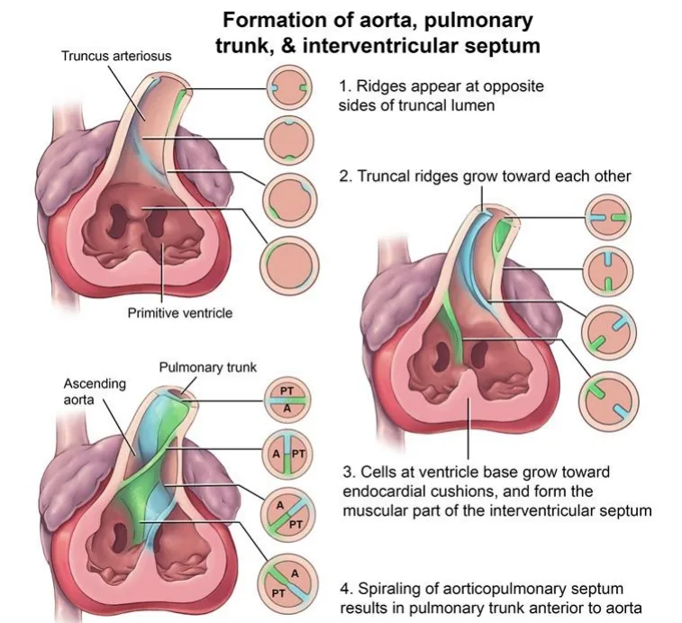
Neural crest cells build the aorticopulmonary septum
they drive the truncal and bulbar ridges to spiral and fuse.- Follow the four stages on the aorta and pulmonary trunk formation figure.
- The spiral is why the pulmonary trunk ends up anterior to the aorta.
Conotruncal defects cluster with DiGeorge syndrome (22q11.2)
because neural crest also builds the pharyngeal pouches.- Look for hypocalcemia, thymic hypoplasia and cleft palate.
| Defect | Mechanism | Page |
|---|---|---|
| Transposition of the great arteries | Aorticopulmonary septum fails to spiral | Transposition of the Great Arteries |
| Tetralogy of Fallot | Anterosuperior displacement of the infundibular septum | Tetralogy of Fallot |
| Persistent truncus arteriosus | Aorticopulmonary septum never forms | Truncus Arteriosus |
Atrial septation
What this shows
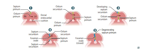
Follow the five panels
on the atrial septation diagram above, and the same sequence inside the heart on the atrial septation anatomy.Septum primum
grows toward the endocardial cushions, narrowing the ostium primum.Ostium secundum
opens within the septum primum.Septum secundum
grows to its right and overlaps the opening.Foramen ovale
the residual channel, valved by the remaining septum primum.At birth the flap closes
left atrial (LA) pressure exceeds right atrial (RA) pressure, so the flap closes and later fuses.- Failure to fuse leaves a Patent Foramen Ovale (~25% of adults).
- It is a flap, not a hole, but still a route for paradoxical embolism.
- A true tissue defect is an Atrial Septal Defect (ASD).
- Secundum ASD is the most common; the primum type belongs to atrioventricular (AV) canal defects and Down syndrome.
Ventricular septation and the AV canal
What this shows
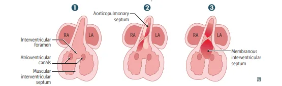
The membranous septum forms last
the muscular septum grows up from the apex, leaving the interventricular foramen.- The aorticopulmonary septum descends and fuses with it to form the membranous septum (three panels on the ventricular septation diagram).
Membranous ventricular septal defect (VSD)
the most common congenital heart defect overall.- Small muscular VSDs usually close on their own (Ventricular Septal Defect).
Endocardial cushions build three things
the atrial septum, the membranous ventricular septum and both AV valves.- That is why a cushion (AV canal) defect gives combined lesions.
- Complete: common AV valve + primum ASD + VSD.
- Partial: primum ASD + cleft mitral valve (mitral regurgitation, MR) with no VSD.
- Strong Down syndrome association.
Valve development
Two sets of cushions
aortic and pulmonary valves come from the outflow-tract cushions; mitral and tricuspid from the fused AV-canal cushions.Four kinds of anomaly
stenotic, regurgitant, atretic (Tricuspid Atresia) or displaced (Ebstein Anomaly, lithium exposure).What each embryonic structure becomes
The organizing rule
smooth walls come from absorbed venous inflow (sinus venosus, pulmonary vein) and the bulbus cordis; trabeculated walls are the original primitive chambers.- The crista terminalis and pectinate muscles mark the boundary.
| Embryonic structure | Gives rise to |
|---|---|
| Truncus arteriosus | Ascending aorta and pulmonary trunk |
| Bulbus cordis | Smooth outflow tracts of both ventricles |
| Primitive ventricle | Trabeculated parts of left ventricle (LV) and right ventricle (RV) |
| Primitive atrium | Trabeculated parts of LA and RA |
| Primitive pulmonary vein | Smooth part of LA |
| Right horn of sinus venosus | Smooth part of RA (sinus venarum) |
| Left horn of sinus venosus | Coronary sinus |
| Right common / anterior cardinal veins | Superior vena cava |
| Endocardial cushions | Atrial septum, membranous ventricular septum, AV and semilunar valves |
Which embryonic structure fails in the combined heart defect of Down syndrome, and what three lesions make up its complete form?
The endocardial cushions, which build the atrial septum, the membranous ventricular septum and both AV valves. The complete AV canal defect is a common AV valve + primum ASD + VSD.
How it's tested
A newborn with hypocalcemic seizures, absent thymic shadow and a conotruncal defect: neural crest failure — DiGeorge (22q11.2 deletion); the cardiac lesion is truncus, tetralogy or an interrupted arch.
Dextrocardia on a chest film in a young adult with chronic sinusitis and infertility: Kartagener syndrome, a dynein defect that also derailed cardiac looping.
A child with Down syndrome has a fixed split S2 and a holosystolic murmur: an endocardial cushion (AV canal) defect — primum ASD plus VSD plus a common AV valve.
Cryptogenic stroke in a 35-year-old with a DVT: paradoxical embolism through a patent foramen ovale — a failure of fusion, present in a quarter of adults.
Go deeper
Related Step 2 pages: Fetal Circulation & Transition at Birth, Approach to Congenital Heart Disease, Atrial Septal Defect, Ventricular Septal Defect, Tetralogy of Fallot, Transposition of the Great Arteries, Truncus Arteriosus
High-yield images4
You just read one page of 236
The rest of Step 2 CK, written exactly like this.
Rapid Review is the reading layer of a full Step 1 platform: a schedule fitted to your exam date, flashcards for every page, and a mentor a message away.
Every Step 1 system
Written the same way: what to know, how it’s tested, where to go deeper.
A plan built to your exam date
Tell us when you sit, and the schedule fits the whole library and your question bank to it.
Flashcards for every page
Ready-made spaced-repetition decks linked to each topic, so nothing you read gets forgotten.
