
Rapid Review·Cardiovascular
Select any text to highlight it or add a note.
Coronary Artery Disease
T1Must knowAcute Coronary Syndrome
Focus on
ECG and troponin sort unstable angina, NSTEMI and STEMI. Everyone gets aspirin, a P2Y12 inhibitor, anticoagulation, a statin and (if safe) a beta blocker. STEMI goes to PCI within 90 minutes or gets lytics within 12 hours; NSTE-ACS gets catheterization within 24 to 72 hours by risk and never fibrinolysis. Then the right ventricular infarct, the stent rules, and the discharge list.
Key takeaways
Three syndromes, one spectrum
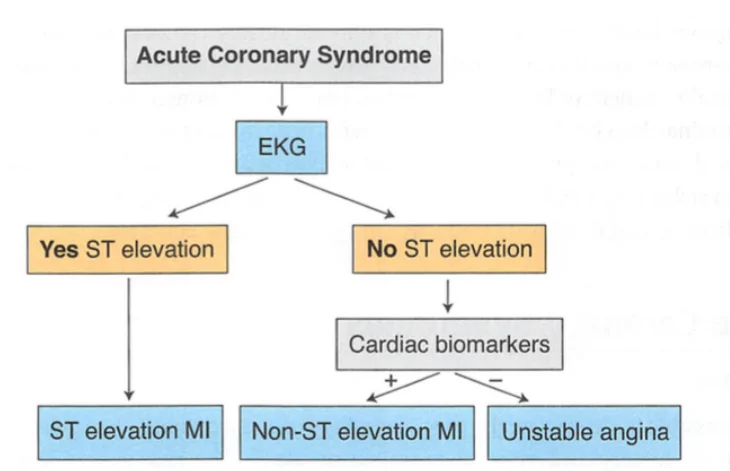
What this shows
Acute coronary syndrome (ACS)
ischemic chest pain lasting over 20 minutes that does not settle with rest; it spans unstable angina and myocardial infarction (MI).- The electrocardiogram (ECG) and troponin split it into three syndromes, as in the ACS table.
Unstable angina
ischemic symptoms with a normal troponin and no ST elevation.Non-ST-elevation myocardial infarction (NSTEMI)
ischemic symptoms with an elevated troponin and no ST elevation.- Unstable angina and NSTEMI share the same ECG picture: ST depression, T-wave inversion, or non-specific changes.
ST-elevation myocardial infarction (STEMI)
ST elevation on the ECG, which means the infarct is transmural.Clinical features
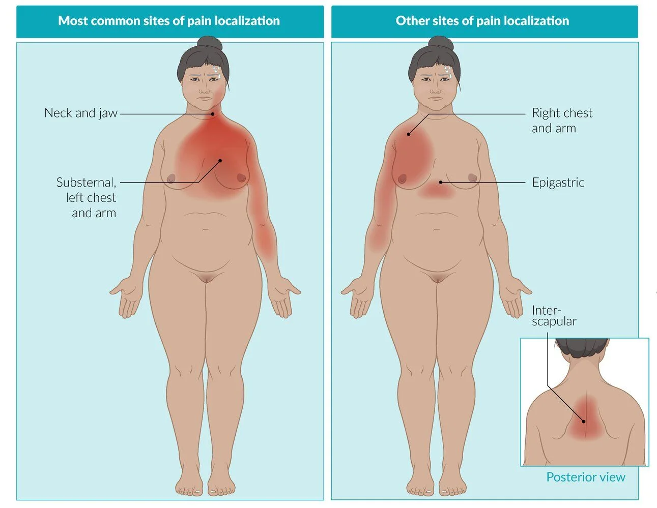
What this shows
Acute retrosternal pressure
dull or squeezing, at rest or with less effort than the patient's usual angina.- It commonly radiates to the arm, shoulder, neck, jaw or epigastrium.
- It may be precipitated by exertion or stress.
Associated symptoms
dyspnea (especially with exertion), pallor, nausea, vomiting, diaphoresis, anxiety (a sense of impending doom), lightheadedness or syncope.Examination
may show tachycardia, an arrhythmia, a new S4, or signs of heart failure or cardiogenic shock.Silent or atypical presentations
in diabetics, women and the elderly, as on the atypical figure.Diagnosis
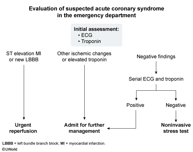
What this shows
ECG within 10 minutes of arrival
it is the gold standard in the first 6 hours.- The first ECG has poor sensitivity, so if it is normal, repeat it every 15 to 30 minutes with serial troponin until the picture resolves or confirms.
- If both stay normal but suspicion is high, order a non-invasive stress test.
STEMI criteria
ST elevation in 2 or more contiguous leads, or a new left bundle branch block (LBBB) with ischemic symptoms (this rules in STEMI even without ST elevation).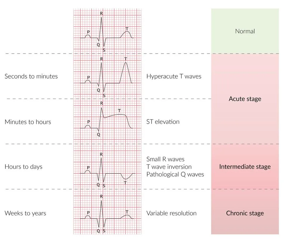
What this shows
ECG timeline
hyperacute T waves come first, then ST elevation.- Pathologic Q waves (persistent, broad and deep) or poor R-wave progression mark an old infarct.
Troponin I
the preferred marker (most sensitive and specific).- A value above the 99th percentile is positive, and it stays elevated for days.
Reinfarction
diagnosed by a troponin rise of 20 percent or more within 3 to 6 hours plus new symptoms or ECG change; a single value cannot do it.- Creatine kinase-MB (CK-MB) was the old reinfarction marker because it normalizes by 48 hours.
Inferior ST elevation mandates right-sided leads (V4R)
to look for right ventricular infarction.Posterior MI
ST depression in V1 to V3 with tall R waves; confirm it with ST elevation in V7 to V9.| Leads | Artery | Wall |
|---|---|---|
| V1 to V2 | Left anterior descending (LAD) | Anteroseptal |
| V3 to V4 | LAD | Anteroapical |
| I, aVL, V5 to V6 | Left circumflex (LCX) | Lateral |
| II, III, aVF | Right coronary artery (RCA) | Inferior |
| Tall R with ST depression in V1 to V3 (ST elevation in V7 to V9) | Posterior descending artery (PDA) | Posterior |
| V4R with inferior changes | RCA | Right ventricle |
Immediate treatment and reperfusion
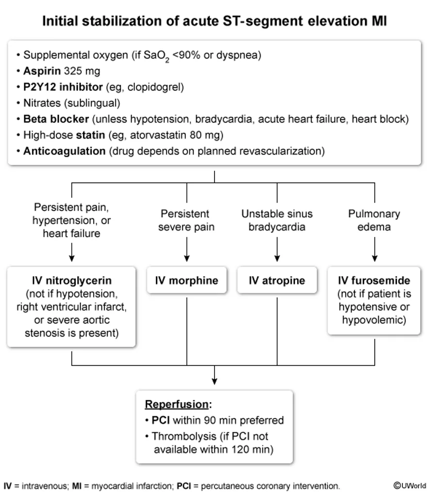
What this shows
Immediate management for everyone (MONA-BASH)
eight parts.- Morphine: only if pain persists.
- Oxygen: only if oxygen saturation (SpO2) is below 90 percent.
- Nitroglycerin: with caution in hypotension (for example right ventricular infarction).
- Antiplatelets: chewed aspirin plus a P2Y12 inhibitor (ticagrelor or clopidogrel) for every patient.
- Beta blocker (oral): contraindicated in cardiogenic shock, hypotension, bradycardia or heart failure.
- Angiotensin-converting enzyme (ACE) inhibitor: within the first 24 hours; it limits post-MI remodeling.
- High-intensity statin.
- Heparin.
Reperfusion (STEMI)
primary percutaneous coronary intervention (PCI) within 90 minutes (120 minutes if the patient must be transferred).- If PCI cannot happen within 120 minutes, give fibrinolysis (tenecteplase or alteplase) within 12 hours of symptom onset.
- Fibrinolysis is contraindicated with a prior intracranial hemorrhage, a recent ischemic stroke, blood pressure above 185/110, or active bleeding.
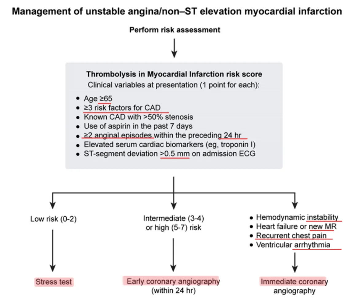
What this shows
Reperfusion (unstable angina and NSTEMI)
risk-stratify with the Thrombolysis in Myocardial Infarction (TIMI) score, then cardiac catheterization (diagnostic and therapeutic) within 24 to 72 hours.- Go immediately to PCI only with shock or hemodynamic instability, ventricular arrhythmia, or refractory chest pain.
- Fibrinolysis is never given for unstable angina or NSTEMI.
Special situations
three that change the plan.- Pulmonary edema: intravenous (IV) furosemide unless the patient is hypotensive, and avoid beta blockers.
- Unstable bradycardia after an inferior MI: IV atropine, then a pacemaker if unresponsive; it is usually transient.
- Heart block after an anterior MI: reflects structural damage, so it will not respond to atropine and needs pacing.
Right ventricular infarction
Usually accompanies an inferior MI
from RCA occlusion.Classic triad
hypotension, raised jugular venous pressure (JVP), and clear lungs.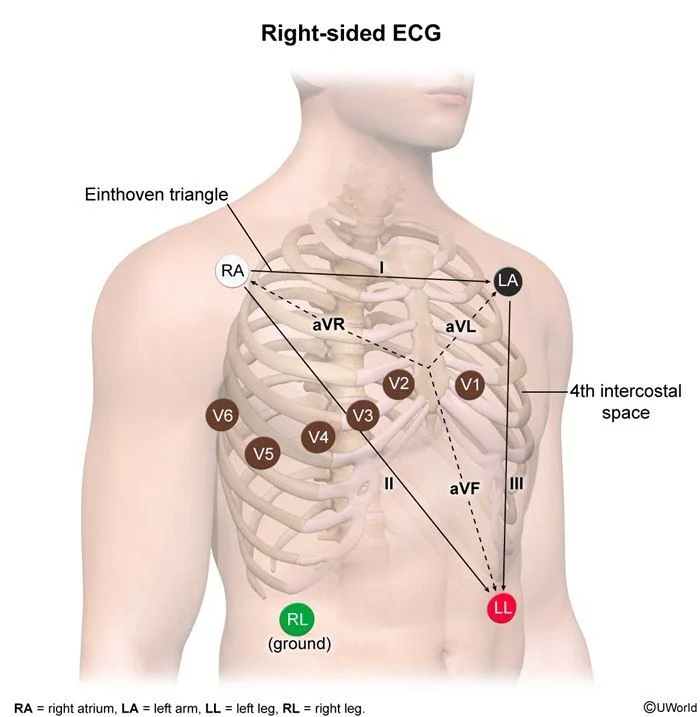
What this shows
Confirm with ST elevation in V4R
the right-sided leads are placed as a mirror image of the usual chest leads.- See also the Read the posterior-inferior STEMI pattern.
Bradycardia and atrioventricular (AV) block are common
because the RCA supplies the AV node.Treat with IV fluids
the right ventricle is preload-dependent.Stents and long-term treatment
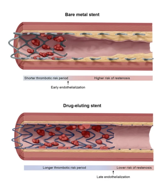
What this shows
An MI after PCI is usually stent thrombosis
acute clot inside the stent, presenting as a STEMI.- Premature discontinuation of antiplatelet therapy is the strongest predictor.
- Treat as a STEMI and go back to PCI.
Bare-metal stents (BMS)
endothelialize early, so their risk is early thrombosis.Drug-eluting stents (DES)
endothelialize late, so their risk is late (over 1 month) and very late (over 1 year) thrombosis.- They need dual antiplatelet therapy (DAPT) for much longer.
In-stent restenosis
angina that returns gradually months after a stent; rare with a DES.Long-term treatment
the goal is secondary prevention.- Continue the antiplatelets, beta blocker, ACE inhibitor or angiotensin receptor blocker (ARB), and high-intensity statin.
- DAPT for 12 months regardless of stent placement, then aspirin for life.
- Add spironolactone when the left ventricular ejection fraction (LVEF) is below 40 percent with heart failure or diabetes.
- Cardiac rehabilitation.
Complications
ventricular fibrillation is the most common cause of death in the first 24 hours.- The mechanical ruptures, aneurysm, pericarditis and Dressler syndrome follow a clock (Post MI Complications).
- Livedo, blue toes and a rising creatinine after catheterization is atheroembolism (Cholesterol Embolism Syndrome).
Inferior ST elevation with hypotension, raised JVP and clear lungs. What else is infarcted, how is it confirmed, and which common ACS drug is dangerous here?
The right ventricle (RCA occlusion). Confirm with ST elevation in V4R on right-sided leads. The right ventricle is preload-dependent, so give IV fluids and avoid nitrates (and morphine and diuretics), which drop preload and cause severe hypotension.
How it's tested
Crushing chest pain, ST elevation in V1 to V4, at a PCI-capable hospital: aspirin, ticagrelor, heparin, and the cath lab within 90 minutes; troponin does not gate this.
Same picture at a rural hospital 3 hours from a cath lab, onset 2 hours ago, no contraindication: fibrinolysis within 30 minutes, then transfer.
Chest pain with ST depression and a positive troponin: NSTEMI; aspirin, P2Y12 inhibitor, heparin, statin, beta blocker; catheterization within 24 hours; fibrinolysis is wrong.
Inferior STEMI with a blood pressure of 80/50, raised neck veins and clear lungs: right ventricular infarction; V4R, IV fluids, no nitrates.
Two months after a DES a man stops clopidogrel before a dental extraction and develops ST elevation in the stented territory: stent thrombosis; emergency catheterization, not a stress test.
Which stent for a patient scheduled for a cancer operation in six weeks: a bare-metal stent or balloon angioplasty, because a DES needs months of uninterrupted DAPT.
Post-MI day 2, EF 30 percent, potassium 4.5, creatinine normal: add eplerenone to the ACE inhibitor and beta blocker; an ICD only if the EF is still 35 percent or less after 40 days.
Go deeper
Guidelines: 2025 ACC/AHA Acute Coronary Syndromes Guideline
Related Step 2 pages: Post MI Complications, Stable Angina, Cholesterol Embolism Syndrome, Cardiogenic Shock, Atrioventricular Block, Heart Failure, Approach to Chest Pain
High-yield images12
Flashcards for this page
Card 1 of 5 · try-out only, nothing is saved
You just read one page of 236
The rest of Step 2 CK, written exactly like this.
Rapid Review is the reading layer of a full Step 1 platform: a schedule fitted to your exam date, flashcards for every page, and a mentor a message away.
Every Step 1 system
Written the same way: what to know, how it’s tested, where to go deeper.
A plan built to your exam date
Tell us when you sit, and the schedule fits the whole library and your question bank to it.
Flashcards for every page
Ready-made spaced-repetition decks linked to each topic, so nothing you read gets forgotten.
