
Rapid Review·Cardiovascular
Select any text to highlight it or add a note.
Coronary Artery Disease
T1Must knowPost MI Complications
Focus on
Timing plus one new finding names the complication: collapse in the first day is ventricular fibrillation; a new murmur at day 3 to 7 is a papillary muscle or septal rupture; sudden pulseless electrical activity at day 5 to 14 is free-wall rupture; persistent ST elevation weeks later is an aneurysm; pleuritic pain weeks later is Dressler syndrome.
Key takeaways
The clock of complications
Three families
complications of myocardial infarction (MI) follow a timeline.- Electrical: first 24 to 48 hours.
- Mechanical: ruptures at 2 to 14 days, when the necrotic wall is softest; aneurysm later.
- Inflammatory or embolic: pericarditis early, Dressler late, mural thrombus.
Early reperfusion is the single best prevention for all of them
late revascularization raises the risk of peri-infarction pericarditis too.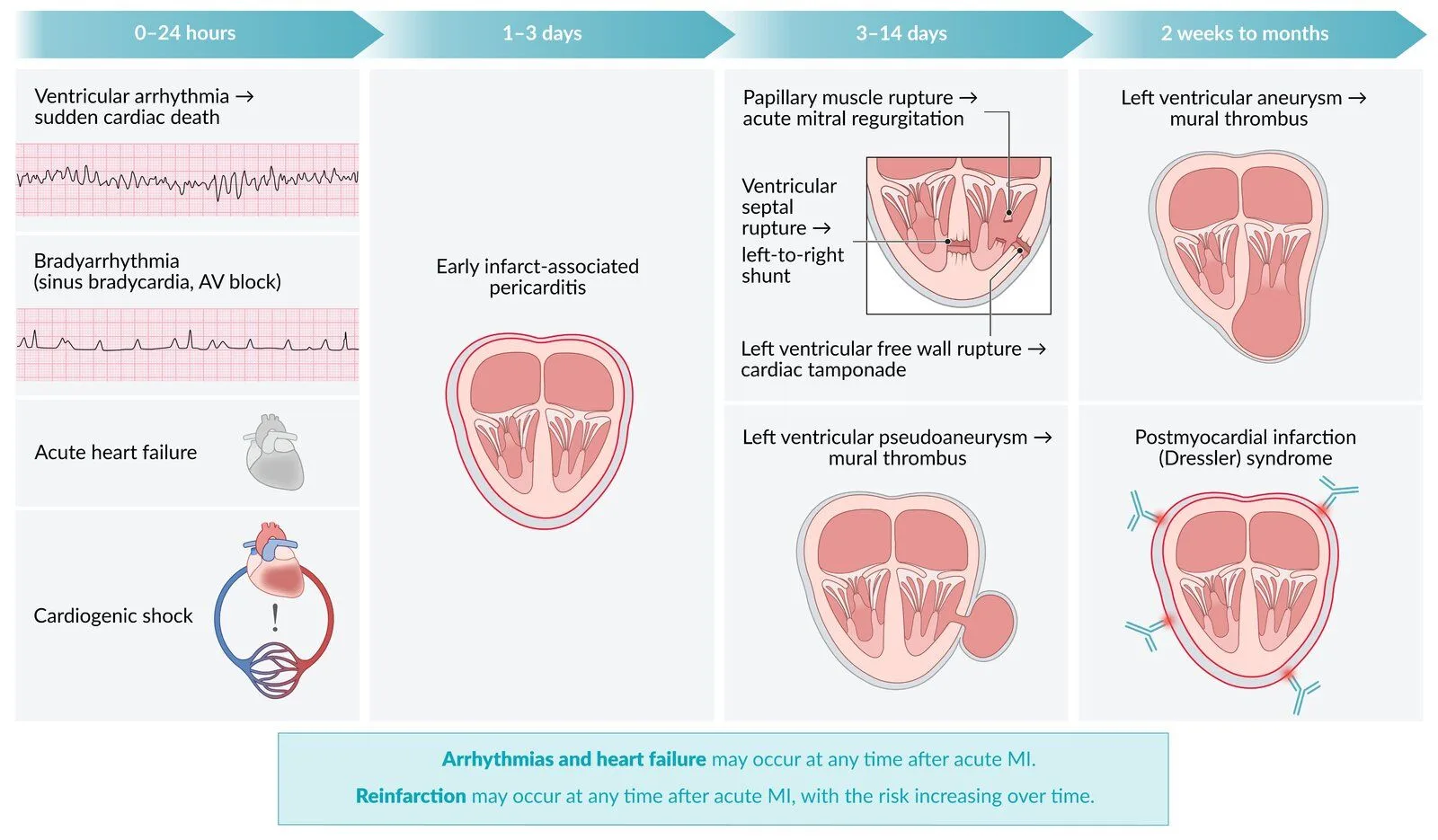
What this shows
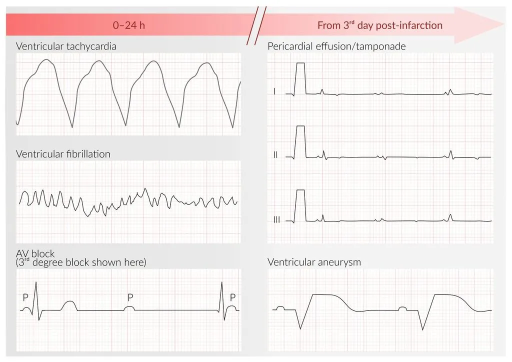
Arrhythmias (mostly under 48 hours)
What this shows
Ventricular fibrillation (VF)
the most common cause of death in the first 24 hours (re-entry in ischemic tissue).- VF is the most common fatal rhythm, ahead of ventricular tachycardia.
- Defibrillate VF or pulseless ventricular tachycardia (VT); amiodarone or lidocaine for recurrent VT; correct potassium and magnesium.
- Early VF does not by itself mandate an implantable cardioverter-defibrillator (ICD); VT or VF beyond 48 hours does.
Bradycardia and atrioventricular (AV) block after inferior MI
the right coronary artery feeds the nodes.- Atropine, then temporary pacing if unstable; usually transient.
Mobitz II or complete block after anterior MI
means septal necrosis.- Pacing, and a worse prognosis.
Mechanical complications
Risk factors
first MI, transmural infarct, late or no reperfusion, older women, hypertension, non-steroidal anti-inflammatory drug (NSAID) or steroid use.All three ruptures present with abrupt deterioration
echocardiography is the diagnostic test.- Surgery is the treatment, with an intra-aortic balloon pump or vasodilators as a bridge.
| Timing | Complication | Clue | Diagnosis and management |
|---|---|---|---|
| 2 to 7 days | Papillary muscle rupture (posteromedial; inferior MI) | Acute pulmonary edema, hypotension, soft apical holosystolic murmur without a thrill; may be nearly silent | Echo shows a flail leaflet and severe mitral regurgitation; afterload reduction, balloon pump, emergency surgery |
| 3 to 5 days | Ventricular septal rupture (apical with anterior MI, basal with inferior MI) | New harsh holosystolic murmur at the left sternal border with a thrill, biventricular failure, oxygen step-up from right atrium to right ventricle | Echo with color Doppler; surgery |
| 5 to 14 days | Free-wall rupture | Sudden hypotension, tamponade, pulseless electrical activity (PEA) arrest | Echo (hemopericardium); pericardiocentesis as a bridge, emergency surgery |
| 3 to 14 days | Pseudoaneurysm | Rupture contained by pericardium; often silent, or murmur, heart failure, syncope | Echo or angiography; urgent surgery (high rupture risk) |
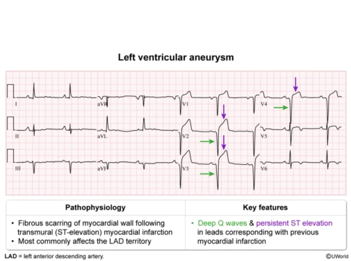
| Weeks to months | True left ventricular aneurysm | Persistent ST elevation, deep Q waves, heart failure, VT, mural thrombus and embolism | Echo (thin dyskinetic wall); ACE inhibitor, anticoagulate if thrombus; surgery rarely |
Pseudoaneurysm vs true aneurysm
a pseudoaneurysm is a free-wall rupture contained by scar and pericardium.- Its risk of rupture into tamponade is higher than a true aneurysm's, which is why it goes to urgent surgery.
Telling the two post-MI murmurs apart
failure pattern and thrill.- Papillary muscle rupture: left-sided failure only (flash pulmonary edema); the murmur is soft or absent because left atrial and ventricular pressures equalize quickly.
- Septal rupture: biventricular failure (pulmonary edema plus a raised jugular venous pressure); the murmur is loud and harsh with a palpable thrill.
Pericarditis (early and late)
Peri-infarction pericarditis (1 to 3 days)
pleuritic positional pain, a rub, diffuse ST elevation over the infarct.- It is an extension of myocardial inflammation, usually localized and self-limiting.
- Treat with high-dose aspirin (for example 975 mg three times a day), with or without acetaminophen.
Dressler syndrome (2 to 10 weeks)
autoimmune pericarditis with fever, malaise, pleuritic pain, a rub, effusion and a raised erythrocyte sedimentation rate (ESR).- Treat with high-dose aspirin (or an NSAID once the infarct has healed) plus colchicine; steroids for refractory cases (Post-Cardiac Injury Syndrome).
Pump failure, thrombus and recovery
Heart failure and cardiogenic shock
follow a large anterior MI (ejection fraction under 40 percent).- Urgent revascularization, inotropes and mechanical support (Cardiogenic Shock), then long-term guideline therapy (Heart Failure).
Left ventricular mural thrombus (anterior MI, aneurysm)
causes embolic stroke, a cold pulseless limb, or renal or mesenteric infarction.- Anticoagulate with warfarin for 3 to 6 months.
What this shows
The aneurysm electrocardiogram (ECG)
persistent ST elevation with deep Q waves in the infarct leads.- True aneurysms also cause angina, heart failure and ventricular arrhythmias.
What this shows
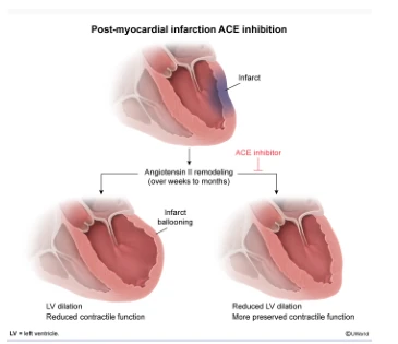
An angiotensin-converting enzyme (ACE) inhibitor follows every large infarct
it limits the remodeling that produces the aneurysm.Recurrent ischemia
new ST elevation in the same territory after a stent is stent thrombosis (Acute Coronary Syndrome).- Livedo, blue toes and a rising creatinine after catheterization is atheroembolism (Cholesterol Embolism Syndrome).
Return to activity
cardiac rehabilitation for everyone.- Moderate exertion and sexual activity resume when the patient can climb two flights of stairs comfortably (typically 1 to 2 weeks after an uncomplicated MI, later after bypass surgery or complications).
- Driving after about a week for an uncomplicated MI.
- Never combine nitrates with phosphodiesterase-5 inhibitors.
Five days after an inferior MI a patient develops sudden pulmonary edema and hypotension with only a soft apical murmur and no thrill. Which complication is this, and how does it differ from a septal rupture?
Papillary muscle rupture (posteromedial, single supply from the posterior descending artery): left-sided failure only, and the murmur is soft because left atrial and ventricular pressures equalize. A septal rupture gives biventricular failure and a loud harsh murmur with a thrill. Both need echo and surgery.
How it's tested
Sudden collapse 6 hours after an ST-elevation myocardial infarction (STEMI); the monitor shows a chaotic rhythm: ventricular fibrillation; defibrillate; it does not by itself mean an ICD.
Day 4 after an inferior MI: sudden dyspnea, crackles, blood pressure 85/50, a soft apical systolic murmur: posteromedial papillary muscle rupture; emergency echo and surgery.
Day 4 after an anterior MI: harsh holosystolic murmur at the left sternal border with a thrill and raised neck veins: ventricular septal rupture.
Day 7: abrupt hypotension, distended neck veins, then PEA: free-wall rupture with tamponade; pericardiocentesis and surgery.
Six weeks after MI: fever, pleuritic chest pain, friction rub, raised ESR: Dressler syndrome; aspirin or NSAID plus colchicine.
Persistent ST elevation with deep Q waves months after an anterior MI, now with a stroke: left ventricular aneurysm with mural thrombus; anticoagulate.
Go deeper
Guidelines: 2025 ACC/AHA Acute Coronary Syndromes Guideline
Related Step 2 pages: Acute Coronary Syndrome, Cardiac Tamponade, Acute Pericarditis, Post-Cardiac Injury Syndrome, Ventricular Tachycardia, Mitral Regurgitation, Cardiac Anatomy
High-yield images4
Flashcards for this page
Card 1 of 5 · try-out only, nothing is saved
You just read one page of 236
The rest of Step 2 CK, written exactly like this.
Rapid Review is the reading layer of a full Step 1 platform: a schedule fitted to your exam date, flashcards for every page, and a mentor a message away.
Every Step 1 system
Written the same way: what to know, how it’s tested, where to go deeper.
A plan built to your exam date
Tell us when you sit, and the schedule fits the whole library and your question bank to it.
Flashcards for every page
Ready-made spaced-repetition decks linked to each topic, so nothing you read gets forgotten.
