
Rapid Review·Cardiovascular
Select any text to highlight it or add a note.
Valvular Heart Disease
T1Must knowMitral Stenosis
Focus on
Almost always rheumatic: a young immigrant woman, often pregnant, with dyspnea, hemoptysis or a stroke from atrial fibrillation, a loud S1, an opening snap and a low-pitched apical diastolic rumble. The closer the snap to S2, the tighter the valve. Balloon valvotomy is the fix; warfarin, not a DOAC, for the AF.
Key takeaways
What it is and what causes it
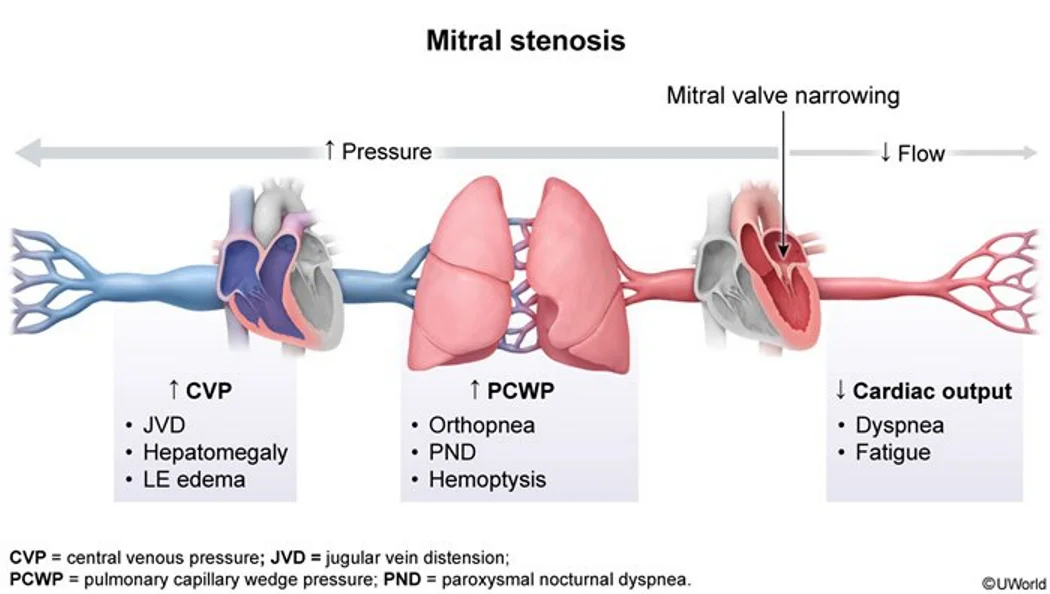
What this shows
Mitral stenosis
obstruction of flow from the left atrium into the left ventricle.- Pressure rises in the left atrium and the pulmonary circulation while left ventricular diastolic pressure stays normal.
Rheumatic heart disease
the most common cause.Senile calcification of the mitral annulus
usually mild.Clinical features
Left heart failure and pulmonary edema
exertional dyspnea, orthopnea, paroxysmal nocturnal dyspnea, hemoptysis; right heart failure follows.Atrial fibrillation
from the dilated atrium, with systemic thromboembolism (stroke).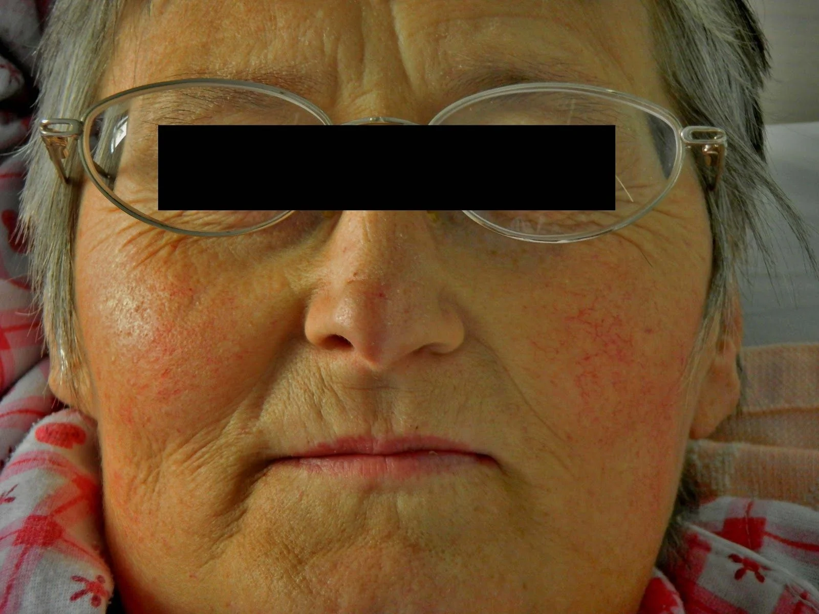
Mitral facies
pink-purple patches on the cheeks.Hoarseness (Ortner syndrome)
the enlarged atrium compresses the recurrent laryngeal nerve.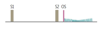
What this shows
Auscultation
an opening snap followed by a mid-diastolic rumble at the apex.- A loud S1 (the stenotic valve snaps shut).
- A loud P2 if pulmonary hypertension has developed.
Diagnosis
Transthoracic echocardiography is the gold standard
thickened, calcified, poorly mobile leaflets, a raised trans-mitral flow velocity, and left atrial enlargement.Severe stenosis
a mitral valve area of 1.5 cm2 or less.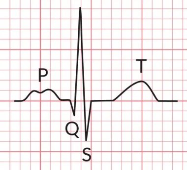
Electrocardiogram
right ventricular hypertrophy (tall R waves in V1 and V2), P mitrale (broad, notched P waves), atrial fibrillation.Chest radiograph
left atrial enlargement and pulmonary congestion.Management
Percutaneous balloon valvotomy
the definitive therapy.- Indicated for severe stenosis (under 1.5 cm2) that is symptomatic, or asymptomatic with pulmonary hypertension.
Medical therapy
for the atrial fibrillation and heart failure.Warfarin
the first-line anticoagulant for valvular atrial fibrillation.A 30-year-old immigrant woman, pregnant, has new orthopnea and hemoptysis, a loud S1, an opening snap soon after S2 and a low-pitched apical diastolic rumble. What is the cause, what is the definitive treatment, and which anticoagulant is used if she develops atrial fibrillation?
Rheumatic mitral stenosis (an early opening snap means a tight valve). The definitive treatment is percutaneous balloon valvotomy. Atrial fibrillation here is treated with warfarin, not a direct oral anticoagulant.
How it's tested
A 32-year-old immigrant at 24 weeks of pregnancy with new orthopnea, a loud S1, an opening snap and an apical diastolic rumble: rheumatic mitral stenosis unmasked by pregnancy — beta blocker, diuretics, and balloon valvotomy if refractory.
Mitral stenosis with new atrial fibrillation — which anticoagulant: warfarin; DOACs are not used in moderate-to-severe rheumatic MS.
Which finding means the stenosis is more severe: an opening snap closer to S2.
Hoarseness and dysphagia in a woman with a diastolic rumble: giant left atrium compressing the recurrent laryngeal nerve and esophagus — Ortner syndrome.
Severe symptomatic MS with a pliable non-calcified valve, no MR, no thrombus: percutaneous balloon valvotomy.
Go deeper
Guidelines: 2020 ACC/AHA Valvular Heart Disease Guideline
Related Step 2 pages: Acute Rheumatic Fever & Rheumatic Heart Disease, Mitral Regurgitation, Atrial Fibrillation, Heart Sounds & Murmurs, Cardiac Tumors, Cardiac Anatomy
High-yield images4
Flashcards for this page
Card 1 of 5 · try-out only, nothing is saved
You just read one page of 236
The rest of Step 2 CK, written exactly like this.
Rapid Review is the reading layer of a full Step 1 platform: a schedule fitted to your exam date, flashcards for every page, and a mentor a message away.
Every Step 1 system
Written the same way: what to know, how it’s tested, where to go deeper.
A plan built to your exam date
Tell us when you sit, and the schedule fits the whole library and your question bank to it.
Flashcards for every page
Ready-made spaced-repetition decks linked to each topic, so nothing you read gets forgotten.
