
Rapid Review·Cardiovascular
Select any text to highlight it or add a note.
Congenital Heart Disease
T1Must knowPatent Ductus Arteriosus
Focus on
A continuous machinery murmur under the left clavicle with bounding pulses and a wide pulse pressure — in a preterm infant, or a term infant exposed to rubella. Indomethacin or ibuprofen close it in the premature; device or surgical closure in everyone else. Never close a duct the baby depends on; and when a large PDA reverses, the cyanosis is in the legs, not the arms.
Key takeaways
What it is and who gets it
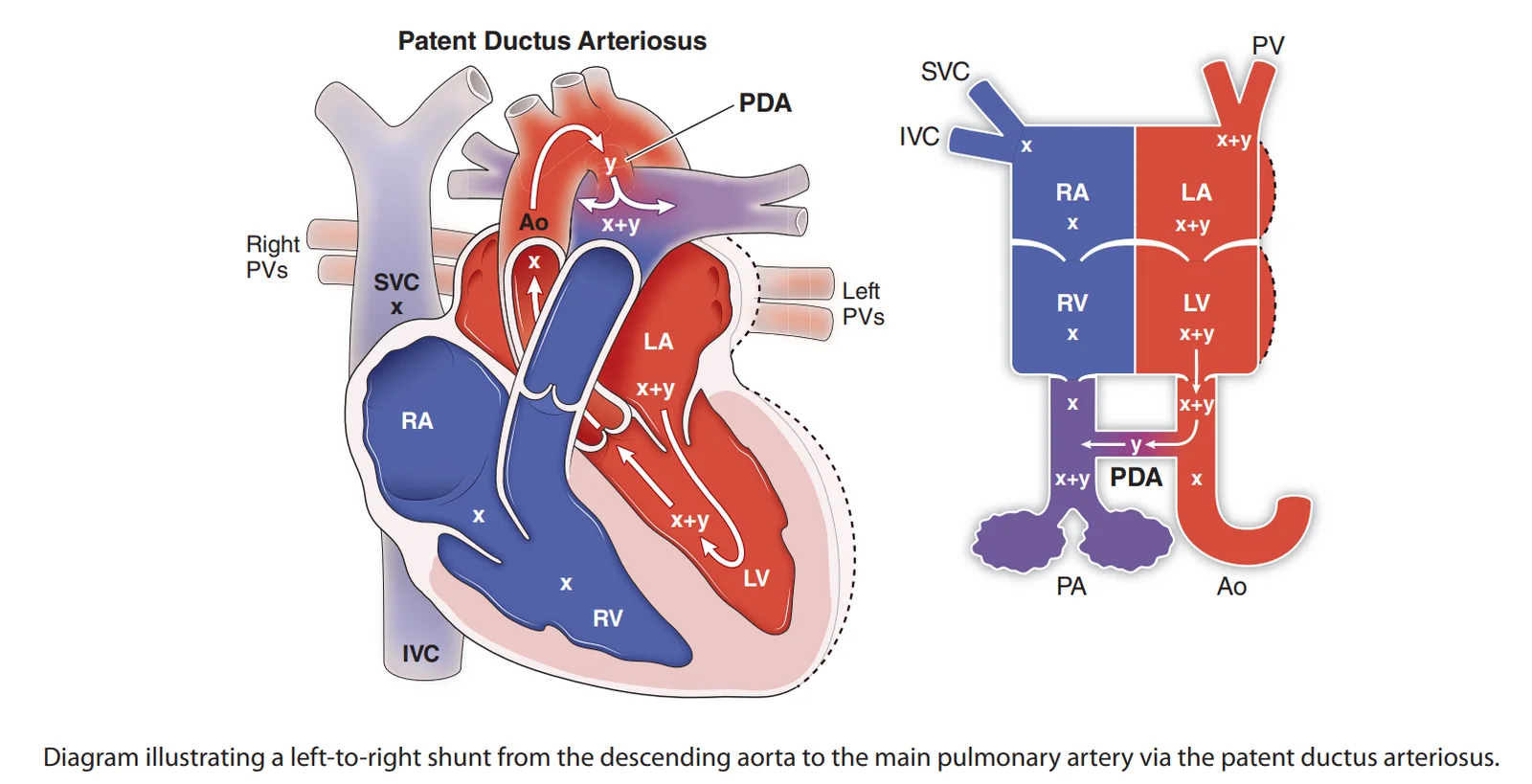
What this shows
Patent ductus arteriosus (PDA)
the ductus normally closes after birth as prostaglandin falls and oxygen saturation rises (Fetal Circulation & Transition at Birth); in a PDA it stays open.The shunt
blood runs from the aorta to the pulmonary artery (left-to-right), over-circulating the lungs and predisposing to pulmonary hypertension.Risk factors
prematurity is the strongest (immature ductal muscle); rubella infection in the first trimester.Clinical features and diagnosis
Small duct
asymptomatic with a normal examination.Large duct
failure to thrive and infant heart failure.- Bounding peripheral pulses and a wide pulse pressure (aortic run-off).
- A loud continuous "machinery" murmur at the left infraclavicular region, loudest at S2.
Shunt reversal
a persistent large shunt reverses with pulmonary hypertension (Eisenmenger Syndrome) and gives differential cyanosis.- Cyanosis of the lower extremities only, because the duct joins the aorta distal to the upper-limb supply.
- Differential cyanosis is also seen in severe preductal coarctation and persistent pulmonary hypertension of the newborn.
Echocardiography is confirmatory
color Doppler shows flow from the aorta into the pulmonary artery; it also excludes a duct-dependent lesion before anyone closes it.Other tests
electrocardiogram, left axis deviation from left ventricular hypertrophy in a large duct; chest film, increased pulmonary markings, a prominent pulmonary artery and aortic knob.| Lesion | Murmur | Site | Other findings |
|---|---|---|---|
| Patent ductus arteriosus | Continuous machinery, peaks at S2 | Left infraclavicular, left upper sternal border | Bounding pulses, wide pulse pressure; differential cyanosis if reversed |
| Ventricular Septal Defect | Harsh holosystolic (smaller is louder) | Left lower sternal border | Hyperdynamic apex; apical rumble when large |
| Atrial Septal Defect | Soft systolic ejection (pulmonic flow) | Left upper sternal border | Wide fixed split S2, right-ventricular heave |
Management and complications
Preterm infants
indomethacin or ibuprofen (prostaglandin synthesis inhibitors; "endomethacin ends the PDA"), or acetaminophen when non-steroidal drugs are contraindicated; surgery if ineffective.Term infants and older
drugs do not work; transcatheter or surgical closure.Complications
heart failure and failure to thrive, pulmonary hypertension and Eisenmenger syndrome with differential cyanosis, infective endarteritis (the reason to close even a small audible duct).- In preterm infants: necrotizing enterocolitis and intraventricular hemorrhage.
A baby born at 28 weeks has bounding pulses, a wide pulse pressure and a continuous machinery murmur below the left clavicle; echo shows aorta-to-pulmonary flow and no duct-dependent lesion. What is the first-line treatment, and what changes in a term infant?
Patent ductus arteriosus in a preterm infant: indomethacin or ibuprofen (acetaminophen if contraindicated), surgery if ineffective. In a term infant drugs do not work, so transcatheter or surgical closure.
How it's tested
A 26-week preterm infant on the ventilator worsens on day 4, with bounding pulses, a wide pulse pressure and a murmur beneath the left clavicle: hemodynamically significant PDA — echocardiogram, then indomethacin or ibuprofen.
A 3-year-old with a continuous murmur at the left upper sternal border, otherwise well: PDA — transcatheter closure (NSAIDs do not work at this age); closing it removes the endocarditis risk.
A teenager with an unrepaired large PDA whose toes are blue and clubbed while the fingers are pink: Eisenmenger syndrome with differential cyanosis — shunt reversal; closure is now contraindicated.
A newborn with hypoplastic left heart syndrome has a large PDA on echo: do not close it — start prostaglandin E1 to keep it open.
What keeps the duct open pharmacologically and what closes it: prostaglandin E1 (alprostadil) keeps it open; indomethacin/ibuprofen (and acetaminophen) close it.
Go deeper
Guidelines: 2018 AHA/ACC Adult Congenital Heart Disease Guideline
Related Step 2 pages: Fetal Circulation & Transition at Birth, Eisenmenger Syndrome, Ventricular Septal Defect, Atrial Septal Defect, Approach to Pediatric Murmurs, Persistent Pulmonary Hypertension of the Newborn
High-yield images1
You just read one page of 236
The rest of Step 2 CK, written exactly like this.
Rapid Review is the reading layer of a full Step 1 platform: a schedule fitted to your exam date, flashcards for every page, and a mentor a message away.
Every Step 1 system
Written the same way: what to know, how it’s tested, where to go deeper.
A plan built to your exam date
Tell us when you sit, and the schedule fits the whole library and your question bank to it.
Flashcards for every page
Ready-made spaced-repetition decks linked to each topic, so nothing you read gets forgotten.
