
Rapid Review·Cardiovascular
Select any text to highlight it or add a note.
Congenital Heart Disease
T1Must knowCoarctation of Aorta
Focus on
Hypertension in the arms, weak delayed pulses in the legs. In the newborn it is shock when the duct closes — prostaglandin E1 first. In the child or young adult it is unexplained hypertension, a systolic murmur over the back, rib notching and a figure-3 sign. Turner syndrome and the bicuspid valve travel with it. Balloon angioplasty or surgical repair, then lifelong blood-pressure surveillance.
Key takeaways
What it is and who gets it
Coarctation of the aorta
thickening of the media of the aortic arch near the ductus narrows the descending aorta, typically just distal to the left subclavian origin.Two pressures
high above the narrowing (arms, head) and low below it (kidneys, legs).- Over years, intercostal collaterals bypass it and notch the ribs.
Associations
Turner syndrome and bicuspid aortic valve (Bicuspid Aortic Valve); an acquired form with Takayasu arteritis; intracranial berry aneurysms.Clinical features
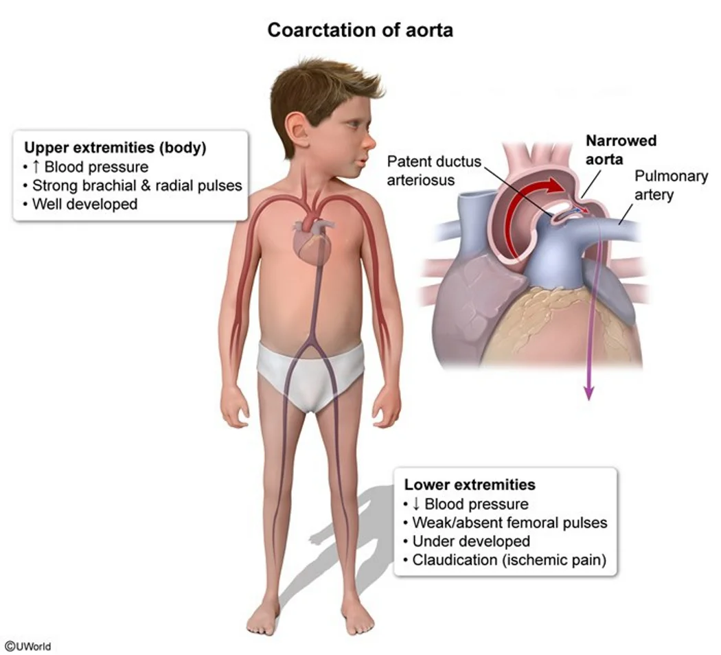
What this shows
Upper-lower body pressure differential
upper-body hypertension (headaches, epistaxis, tinnitus) with strong arm pulses and a well-developed upper body.- Weak femoral pulses with brachial-femoral delay, lower leg pressure and a poorly developed lower body.
- Lower postductal saturation (differential cyanosis).
Neonates with severe narrowing
heart failure (irritability, poor feeding, diaphoresis) and cardiogenic shock a few days after birth when the ductus closes.Children and adults (mild narrowing)
cold legs with claudication on exercise; palpable intercostal collateral pulsations in adults.Auscultation
two murmurs.- A systolic ejection murmur over the left posterior hemithorax (flow through the narrowing).
- A continuous murmur in the left infraclavicular and interscapular region (collateral flow).
Diagnosis
Upper and lower extremity blood pressure is the best initial test
arm pressure higher than leg pressure.- If the left subclavian origin is involved, the right arm reads higher than the left.
Echocardiography is confirmatory
it assesses severity, as drawn on the Review the chest radiograph and ductal-flow anatomy.- Computed tomography (CT) or magnetic resonance (MR) angiography defines the arch and collaterals in older patients.
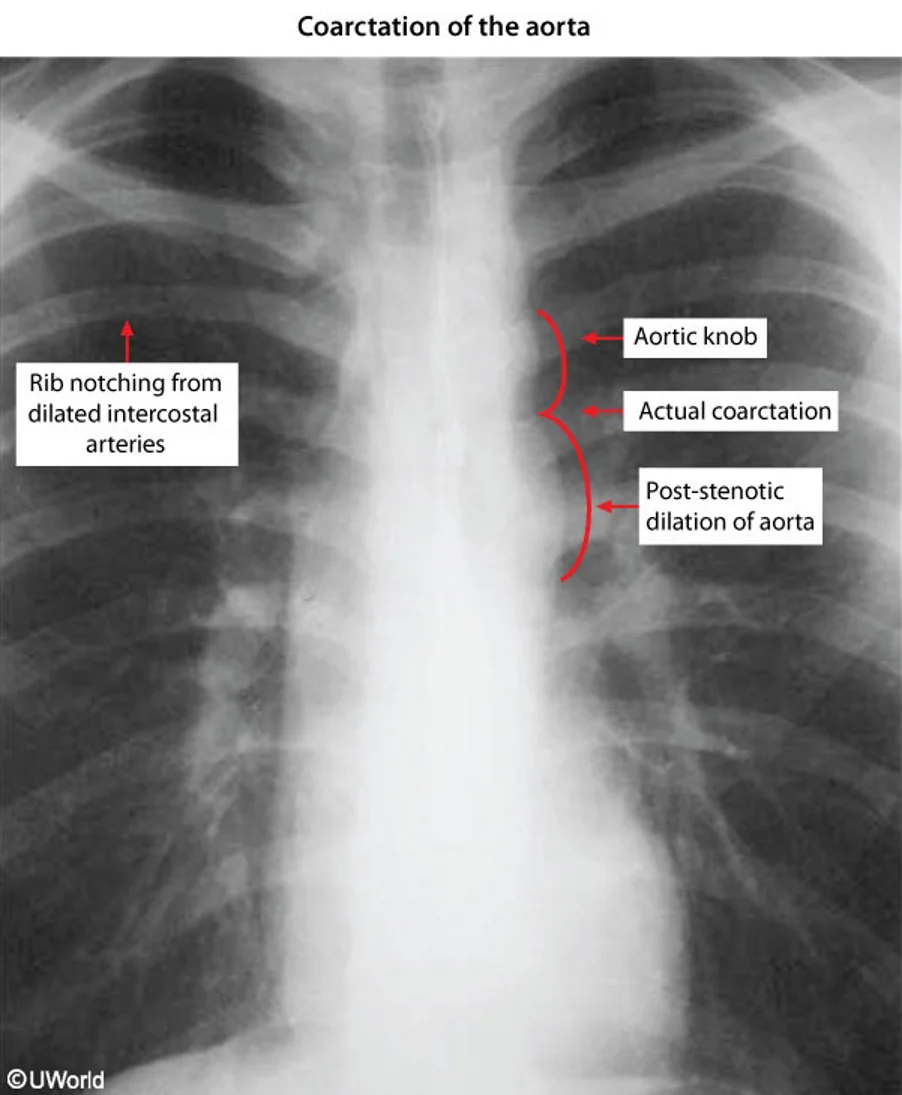
What this shows
Chest film
the figure-3 sign (an indentation at the coarctation with post-stenotic dilation) and inferior notching of the 3rd to 8th ribs from intercostal collaterals, seen after about 5 years of age, not in infants.Other tests
pulse oximetry shows a lower post-coarctation saturation; the electrocardiogram shows left ventricular hypertrophy.| Critical (neonatal) coarctation | Childhood or adult coarctation | |
|---|---|---|
| Timing | Days 3 to 14, as the ductus closes | Any age; often found with hypertension |
| Picture | Heart failure, shock, acidosis, absent femoral pulses, differential cyanosis | Upper-limb hypertension, brachial-femoral delay, leg claudication, interscapular murmur |
| Film | Cardiomegaly, pulmonary edema | Figure-3 sign, rib notching (over 5 years) |
| First step | Prostaglandin E1, stabilize, then repair | Confirm with echo or CT, then balloon angioplasty or surgery |
Management and complications
Newborns with suspected severe coarctation
prostaglandin E1 immediately to maintain the ductus, then stabilize and repair.Significant stenosis
surgical correction or balloon angioplasty (with stenting in older children and adults, and for recurrence after surgery).Long term
antihypertensives before and often after repair; lifelong follow-up of the repair site, the bicuspid valve and the blood pressure.Complications
aortic aneurysm, dissection and rupture; cerebral berry aneurysms and subarachnoid hemorrhage; hypertension with left ventricular hypertrophy and heart failure; endocarditis; recoarctation after repair.A 16-year-old girl with short stature and a webbed neck has an arm blood pressure of 160/90, weak delayed femoral pulses and a murmur between the scapulae; the film shows notched ribs. What is the diagnosis, the best initial test, and a key associated valve lesion?
Coarctation of the aorta in Turner syndrome. The best initial test is upper and lower extremity blood pressure; echo confirms. Look for a bicuspid aortic valve (and berry aneurysms); treat with surgery or balloon angioplasty with stenting.
How it's tested
A 10-day-old previously well newborn is grey, tachypneic and acidotic with absent femoral pulses and a saturation of 92% in the hand but 78% in the foot: critical coarctation with ductal closure — prostaglandin E1 now, then echo and surgical repair.
A 14-year-old with BP 160/95 in the arms and 100/60 in the legs, a delayed femoral pulse and a murmur heard best between the scapulae: coarctation — echocardiogram and CT angiography; balloon angioplasty with stent.
Rib notching on the film of a teenager with unexplained hypertension: dilated intercostal collaterals from coarctation — the notches take years to form, so this is not a neonatal finding.
A short girl with a webbed neck, hypertension and an ejection click: Turner syndrome — coarctation plus bicuspid aortic valve.
Sudden severe headache in a 30-year-old with repaired coarctation: ruptured berry aneurysm — CT head; coarctation patients carry the association for life.
Go deeper
Guidelines: 2018 AHA/ACC Adult Congenital Heart Disease Guideline
Related Step 2 pages: Approach to Congenital Heart Disease, Approach to Neonatal Cyanosis, Bicuspid Aortic Valve, Secondary Hypertension, Fetal Circulation & Transition at Birth, Hypoplastic Left Heart Syndrome
High-yield images3
Flashcards for this page
Card 1 of 5 · try-out only, nothing is saved
You just read one page of 236
The rest of Step 2 CK, written exactly like this.
Rapid Review is the reading layer of a full Step 1 platform: a schedule fitted to your exam date, flashcards for every page, and a mentor a message away.
Every Step 1 system
Written the same way: what to know, how it’s tested, where to go deeper.
A plan built to your exam date
Tell us when you sit, and the schedule fits the whole library and your question bank to it.
Flashcards for every page
Ready-made spaced-repetition decks linked to each topic, so nothing you read gets forgotten.
