
Rapid Review·Cardiovascular
Select any text to highlight it or add a note.
Cardiomyopathy
T1Must knowRestrictive Cardiomyopathy
Focus on
Stiff, infiltrated walls with a normal EF and right-sided congestion. Amyloid is the answer when the walls are thick but the ECG voltage is low; sarcoid when there is heart block; hemochromatosis when there is diabetes and bronze skin. The exam's favorite twin is constrictive pericarditis — which is curable.
Key takeaways
What it is and what causes it
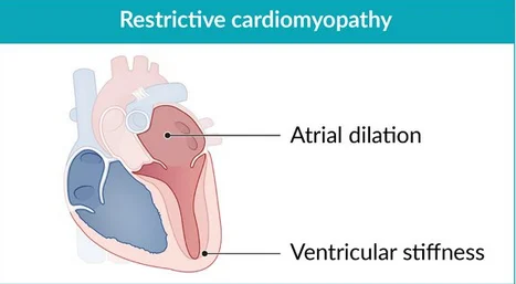
What this shows
An infiltrating process stiffens the muscle
a process infiltrates the myocardium and impairs diastolic relaxation and filling.- Systolic function is spared, so the ejection fraction is normal or near-normal.
Infiltrative disease
the main group.- Amyloidosis (the likely answer when the wall is markedly thickened).
- Sarcoidosis (conduction abnormalities).
- Hemochromatosis (restrictive early, dilated late).
- Fabry disease, scleroderma.
In young children
endocardial fibroelastosis, and Loeffler syndrome (endomyocardial fibrosis with an eosinophilic infiltrate on histology).Radiation
through scarring and fibrosis.Clinical features
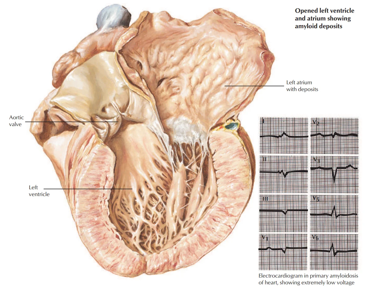
What this shows
Diastolic heart failure with right-sided symptoms predominating
jugular venous distension, peripheral edema, ascites.Kussmaul sign
the neck veins rise on inspiration.Diagnosis
Transthoracic echocardiography
the gold standard.- A restrictive filling pattern with bi-atrial enlargement and a normal to near-normal ejection fraction.
- Early diastolic filling is normal; late diastolic filling is halted.
Electrocardiogram (ECG)
low voltage (especially amyloidosis), sometimes a left bundle branch block.Endomyocardial biopsy
sometimes used to confirm the cause.Management
Treat like heart failure with preserved ejection fraction
(Heart Failure) diuretics are first line.Treat the underlying cause
when possible.Severe or refractory symptoms
consider heart transplant.A patient has prominent jugular venous distension that rises on inspiration, ascites and edema; echo shows thick walls, dilated atria and a normal ejection fraction, and the ECG shows low voltage. What is the likely cause and the first-line treatment?
Restrictive cardiomyopathy from amyloidosis: markedly thickened walls with low voltage, bi-atrial enlargement and a preserved ejection fraction, with right-sided signs and Kussmaul sign. Treat like heart failure with preserved ejection fraction: diuretics first, and treat the cause.
How it's tested
A 70-year-old man with heart failure, thick LV walls on echo, low voltage on ECG, proteinuria and bilateral carpal tunnel syndrome: cardiac amyloidosis — PYP scan and light chains; diuretics; avoid digoxin and CCBs.
Young adult with heart failure, complete heart block and hilar lymphadenopathy: cardiac sarcoidosis — glucocorticoids and a pacemaker/ICD.
Right heart failure with a pericardial knock and calcified pericardium after tuberculosis: constrictive pericarditis, not restriction — pericardiectomy cures it.
Heart failure with diabetes, bronze skin and a ferritin of 2,000: hemochromatosis — phlebotomy.
Go deeper
Guidelines: 2023 ESC Cardiomyopathy Guideline
Related Step 2 pages: Dilated Cardiomyopathy, Heart Failure, Constrictive Pericarditis, Jugular Venous Pressure, Atrioventricular Block
High-yield images2
You just read one page of 236
The rest of Step 2 CK, written exactly like this.
Rapid Review is the reading layer of a full Step 1 platform: a schedule fitted to your exam date, flashcards for every page, and a mentor a message away.
Every Step 1 system
Written the same way: what to know, how it’s tested, where to go deeper.
A plan built to your exam date
Tell us when you sit, and the schedule fits the whole library and your question bank to it.
Flashcards for every page
Ready-made spaced-repetition decks linked to each topic, so nothing you read gets forgotten.
