
Rapid Review·Cardiovascular
Select any text to highlight it or add a note.
Cardiomyopathy
T1Must knowHypertrophic Cardiomyopathy
Focus on
The young athlete with exertional syncope, a family history of sudden death and a systolic murmur that gets louder on standing. Confirm with echo (septal hypertrophy, SAM, outflow gradient), treat with a beta blocker, avoid everything that shrinks the ventricle, and assess for an ICD. Know how to tell it from an athlete's heart and from aortic stenosis.
Key takeaways
What it is and what causes it
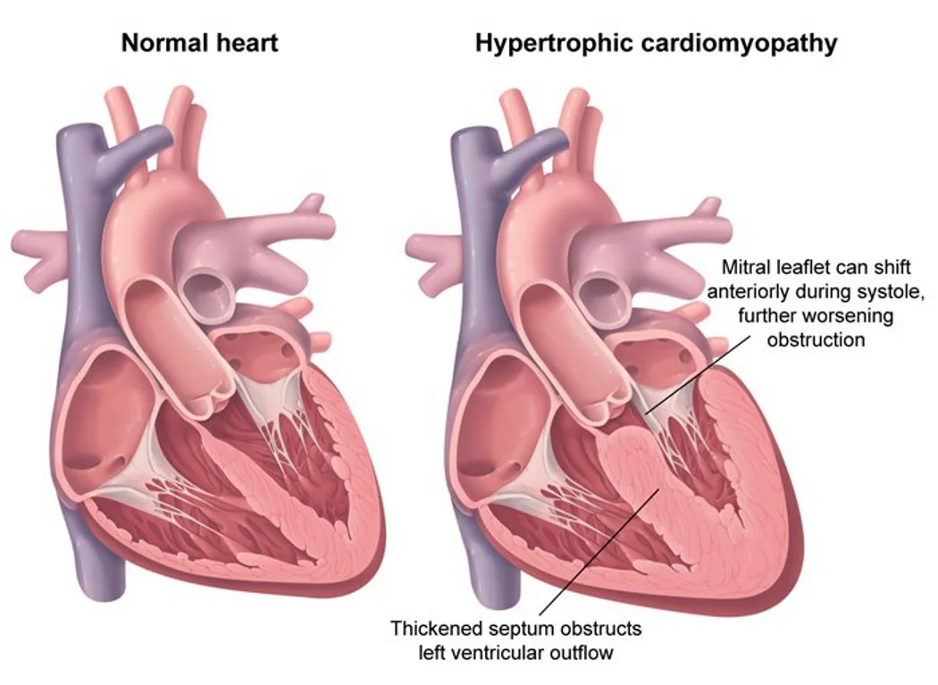
What this shows
Hypertrophic cardiomyopathy (HCM)
a disorder of sarcomere proteins with disorganized, hypertrophied myocytes.- It causes diastolic dysfunction (impaired relaxation and filling).
- With concentric hypertrophy: sarcomeres are added in parallel, so the walls thicken.
Hypertrophic obstructive cardiomyopathy (HOCM)
the congenital variant with predominant septal hypertrophy that obstructs the left ventricular outflow tract (LVOT).- See the HCM anatomy and management pathway.
Congenital HOCM
usually an autosomal dominant mutation in beta-myosin heavy chain or myosin-binding protein C.- Also Friedreich ataxia, Fabry disease and Noonan syndrome.
Acquired
hypertension (the most common acquired cause) and aortic stenosis.Clinical features
Presentation
exertional syncope, chest pain, dyspnea from heart failure, or sudden cardiac death.- Syncope is multifactorial: outflow obstruction, arrhythmia, ischemia, or an inappropriate baroreceptor-driven vasodilation.
Rapid carotid upstroke
blood leaves quickly in early systole before the outflow tract collapses in mid-to-late systole.Auscultation
an S4 and a harsh systolic ejection murmur at the left lower sternal border, with or without mitral regurgitation from impaired mitral closure.Arrhythmia risk
HCM raises the risk of atrial fibrillation (dilated left atrium) and ventricular tachycardia.- So periodic ambulatory ECG monitoring is indicated.
Diagnosis
Transthoracic echocardiography (TTE)
the structural diagnosis.- Asymmetric septal hypertrophy with a wall over 15 mm (normal under 11 mm).
- Dynamic LVOT obstruction and diastolic filling dysfunction.
- A dilated left atrium with a small left ventricular cavity.
- Systolic anterior motion (SAM) of the mitral valve.
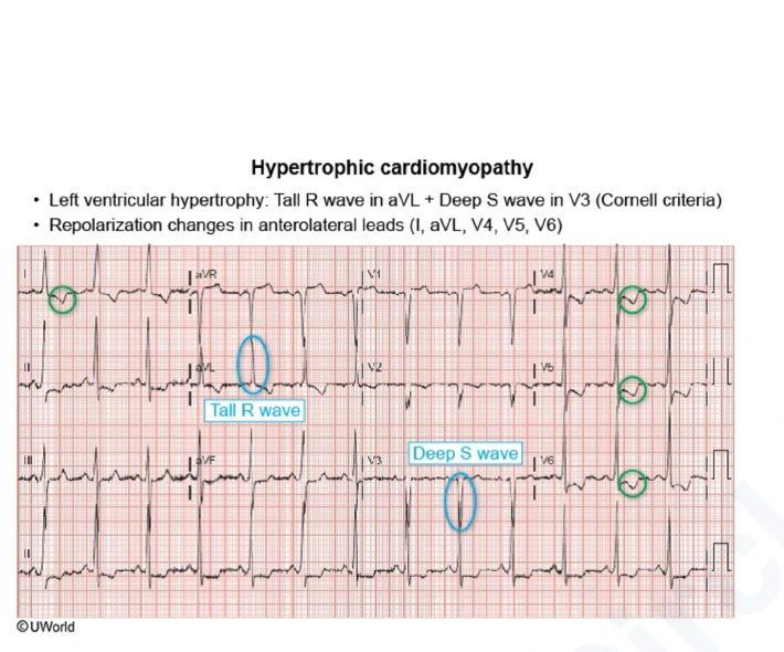
What this shows
Electrocardiogram (ECG)
left ventricular hypertrophy (LVH) with left axis deviation (tall R in aVL plus deep S in V3, the Cornell criteria), prominent Q waves, and T-wave inversions.Management and ICD
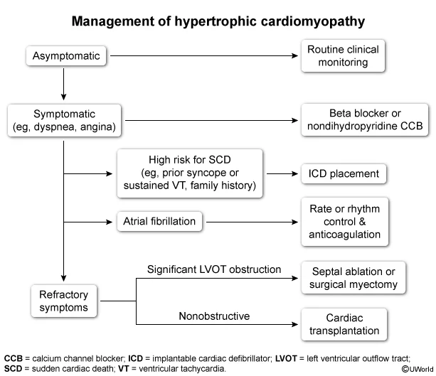
What this shows
Asymptomatic
avoid strenuous exercise.Symptomatic
beta blockers first line; a calcium-channel blocker, especially verapamil, is second line.- The goal is negative inotropy and chronotropy to lengthen diastolic filling.
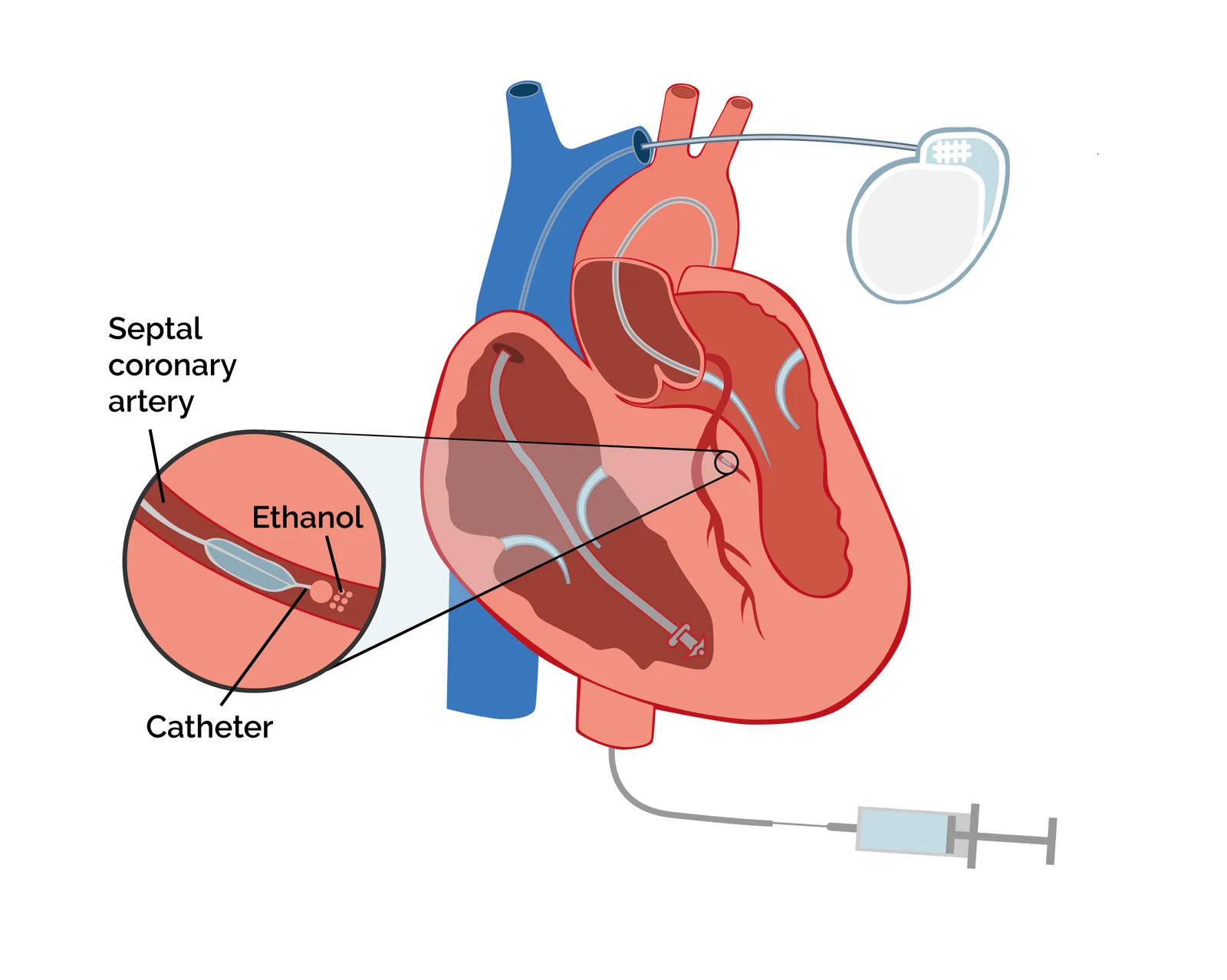
What this shows
Persistent symptoms despite drugs
septal myectomy or alcohol septal ablation.Implantable cardioverter-defibrillator (ICD), secondary prevention
the best way to prevent sudden death, after sudden cardiac arrest or sustained ventricular tachycardia.ICD, primary prevention
a list of risk markers.- Sudden cardiac death in a close relative under 50.
- Unexplained or exertional syncope.
- A wall thickness of 30 mm or more.
- Non-sustained ventricular tachycardia on Holter, an apical aneurysm or an ejection fraction under 50 percent, or a hypotensive blood pressure response to exercise.
Prognosis, infants and athletes
Prognosis
most patients have a normal life expectancy with treatment; no symptoms and diagnosis in adulthood are favorable.- The risk of sudden death falls with age and is greatest under 30.
- Poor prognosis: diagnosis in infancy or early childhood, wall thickness over 25 mm, an outflow gradient over 30 mm Hg, and especially the development of systolic dysfunction; women do slightly worse than men.
What this shows
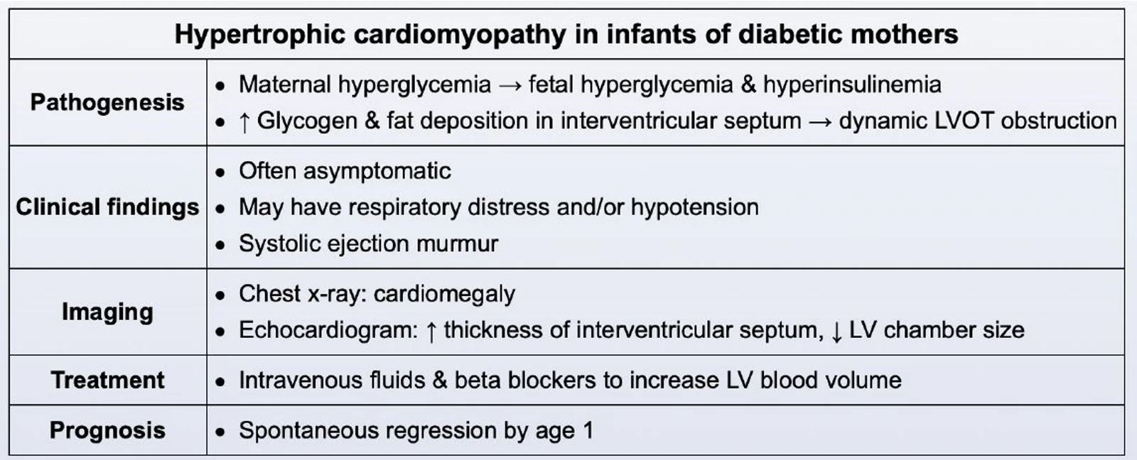
HCM in the infant of a diabetic mother
maternal hyperglycemia causes fetal hyperinsulinemia, and insulin drives glycogen and fat deposition in the septum, giving dynamic LVOT obstruction.- Often asymptomatic, or heart failure with respiratory distress, tachycardia or hypotension, and a systolic ejection murmur.
- Chest radiograph shows cardiomegaly; echo shows a thick septum and a small left ventricular cavity.
- Treat with intravenous fluids and beta blockers to increase left ventricular volume; it regresses spontaneously by age 1.
What this shows
HCM vs athlete's heart
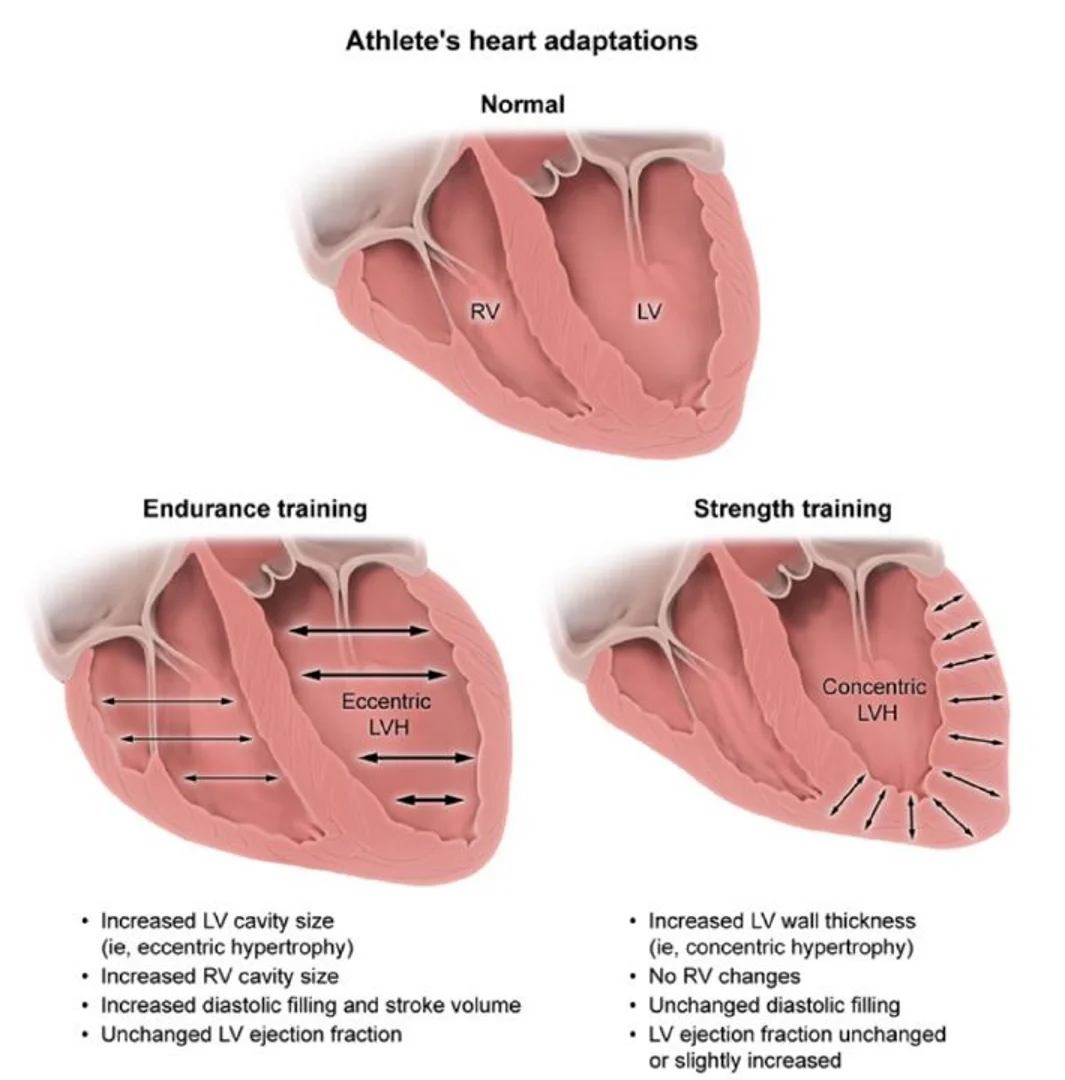
training thickens the heart too, but in a balanced way.- Endurance training: larger ventricular cavities (eccentric LVH), better filling.
- Strength training: thicker walls (concentric LVH), with normal diastolic filling.
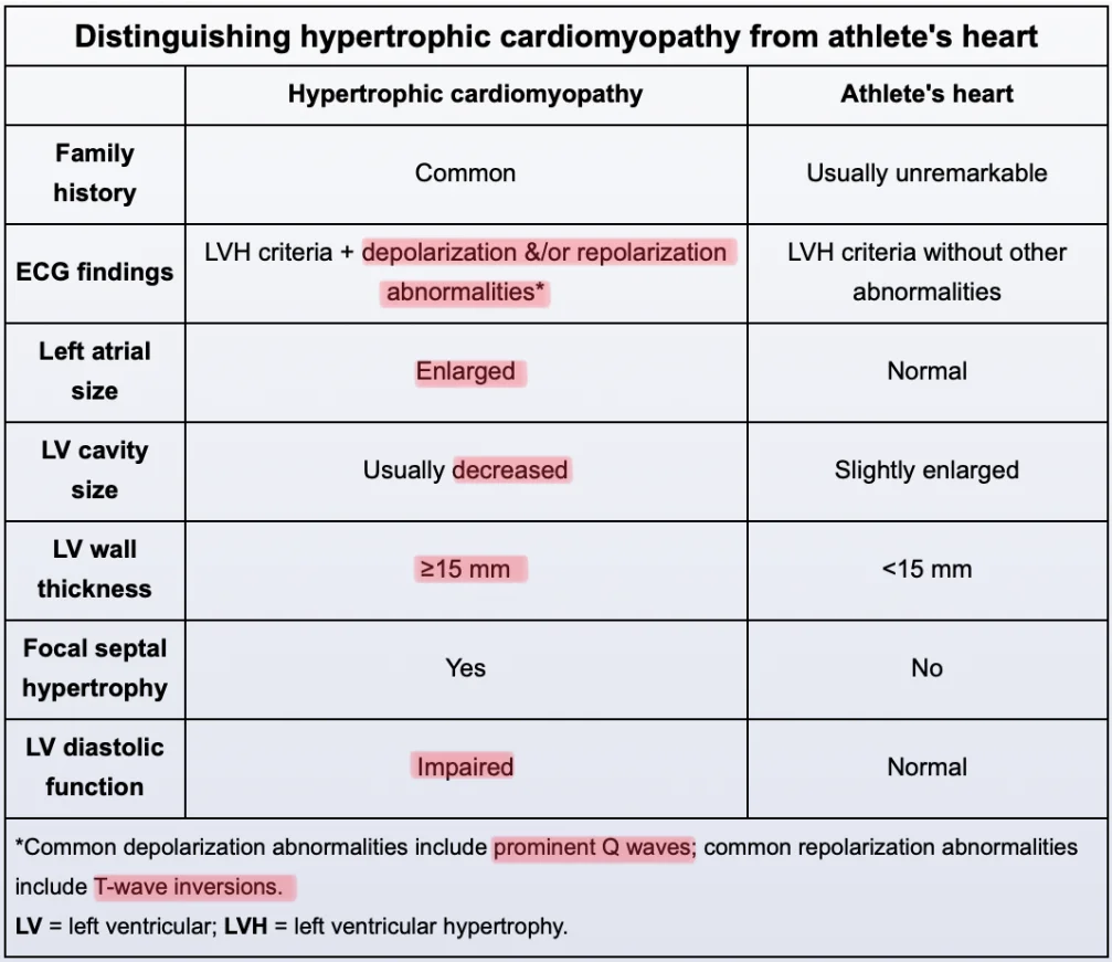
- An isolated T-wave inversion in V1 is common in athletes; T-wave inversions in 2 contiguous leads point to HCM.
| Feature | Hypertrophic cardiomyopathy | Athlete's heart |
|---|---|---|
| Family history | Common | Usually unremarkable |
| ECG | LVH plus depolarization or repolarization abnormalities (prominent Q waves, T-wave inversions) | LVH voltage only |
| Left atrium | Enlarged | Normal |
| Left ventricular cavity | Usually small | Slightly enlarged |
| Wall thickness | 15 mm or more | Under 15 mm |
| Focal septal hypertrophy | Yes | No |
| Diastolic function | Impaired | Normal |
What this shows
A young athlete has a harsh systolic murmur at the left lower sternal border that gets louder when standing. Echo shows a 19 mm septum, a small left ventricular cavity and systolic anterior motion of the mitral valve. What is it, what is the first-line drug, and which drugs must be avoided?
Hypertrophic obstructive cardiomyopathy (wall over 15 mm, small cavity and septal hypertrophy separate it from athlete's heart). First line is a beta blocker (verapamil second). Avoid digoxin, nitrates, dihydropyridine calcium-channel blockers, ACE inhibitors and diuretics, which shrink the ventricle and worsen the obstruction.
How it's tested
A 17-year-old collapses during basketball; his uncle died suddenly at 30; a left sternal border murmur gets louder with Valsalva: hypertrophic cardiomyopathy — echocardiogram, then ICD evaluation.
HOCM patient given nitroglycerin for chest pain becomes hypotensive: preload fell and the obstruction worsened — fluids, stop the vasodilator.
Symptomatic HOCM without shock — first drug: a beta blocker; verapamil if not tolerated.
HCM with new AF and a CHA2DS2-VASc of 0: anticoagulate anyway.
A murmur that radiates to the carotids with a slow weak pulse and softens on Valsalva: aortic stenosis, not HCM.
A newborn of a poorly controlled diabetic mother has a thick septum on echo: transient hypertrophy — supportive care, resolves.
Go deeper
Guidelines: 2024 AHA/ACC HCM Guideline · 2023 ESC Cardiomyopathy Guideline
Related Step 2 pages: Dilated Cardiomyopathy, Heart Sounds & Murmurs, Aortic Stenosis, Approach to Syncope, Atrial Fibrillation, Cardiac Implantable Devices, Anomalous Aortic Origin of a Coronary Artery
High-yield images8
Flashcards for this page
Card 1 of 5 · try-out only, nothing is saved
You just read one page of 236
The rest of Step 2 CK, written exactly like this.
Rapid Review is the reading layer of a full Step 1 platform: a schedule fitted to your exam date, flashcards for every page, and a mentor a message away.
Every Step 1 system
Written the same way: what to know, how it’s tested, where to go deeper.
A plan built to your exam date
Tell us when you sit, and the schedule fits the whole library and your question bank to it.
Flashcards for every page
Ready-made spaced-repetition decks linked to each topic, so nothing you read gets forgotten.
