
Rapid Review·Cardiovascular
Select any text to highlight it or add a note.
Coronary Artery Disease
T1Must knowStable Angina
Focus on
Predictable chest pressure on exertion from a fixed coronary plaque. Confirm it with pretest probability and a stress test, relieve attacks with nitroglycerin, prevent them with a beta blocker, and protect life with aspirin, a statin and risk-factor control. Revascularize only for refractory symptoms or high-risk anatomy.
Key takeaways
What it is and who gets it
Coronary artery disease (CAD)
ischemic heart disease caused by atherosclerotic change in the coronary arteries.- It is generally silent until the plaque narrows the lumen by more than 75 percent.
Stable angina is the symptom of a fixed plaque
on exertion, oxygen demand exceeds what the narrowed artery can supply.- Plaque rupture with thrombus is a different disease (Acute Coronary Syndrome).
- Spasm without plaque is Vasospastic Angina.
Smoking is the strongest risk factor
the others add up.- Obesity, dyslipidemia, hypertension, diabetes, chronic kidney disease (CKD).
- Male sex, and age over 45 in men or over 55 (or post-menopause) in women.
- Family history of premature CAD: a first-degree relative with CAD before 55 (male) or before 65 (female).
CAD equivalents are treated as if CAD is already present
diabetes mellitus, CKD, and non-coronary atherosclerosis (Peripheral Artery Disease, Carotid Artery Stenosis, Abdominal Aortic Aneurysm).Clinical features
Exertional chest pain
comes on with exertion and is relieved by rest or nitroglycerin.- Pain can show in NBME as feeling pressure on the chest.
Three criteria define typical angina
count how many the pain meets.- Location and quality: substernal, diffuse pressure or squeezing.
- Trigger: brought on by exertion or intense emotion.
- Relief: relieved by rest or nitroglycerin.
- Typical angina meets all three, atypical meets two, and non-cardiac chest pain meets one or none.
Atypical presentation
diabetics, the elderly and women may have silent ischemia.- In acute coronary syndrome (ACS) the pain may be absent or replaced by autonomic symptoms (nausea, diaphoresis).
- Silent angina is also seen in patients with frequent repeated attacks.
Diagnosis and pretest probability
The diagnosis is clinical
classic symptoms plus risk factors.Resting electrocardiogram (ECG) first
it is the best initial test and is done for every symptomatic patient.- It is usually normal or non-specific between attacks.
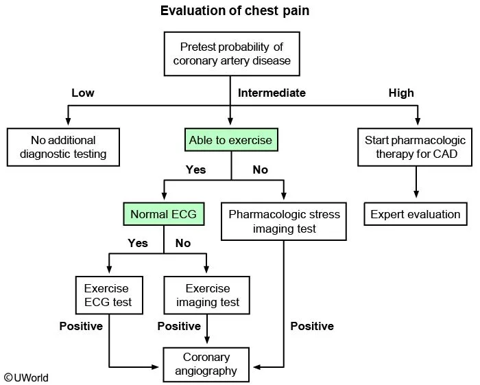
What this shows
Then estimate the pretest probability
from age, sex and how typical the pain is.| Probability | Clinical scenario | Next step |
|---|---|---|
| Low | Asymptomatic people of any age; atypical chest pain in a woman under 50 | No additional testing |
| Intermediate | Men with atypical angina (any age); women with atypical angina over 50; women with typical angina aged 30 to 50 | Stress test, or coronary computed tomography angiography (CCTA) |
| High | Typical angina in men aged 40 or more, or women aged 60 or more | Start pharmacologic therapy for CAD |
A positive stress test or CCTA leads to invasive coronary angiography
the two are different tests.- CCTA is non-invasive.
- Coronary angiography is the invasive digital subtraction study, and it is the route to revascularization.
Stress testing
Which stress test to perform
it depends on whether the patient can exercise.- Exercise ECG is first choice if the patient can exercise; echocardiography or nuclear perfusion imaging can be added.
- Pharmacologic stress if the patient cannot exercise: a vasodilator (dipyridamole, adenosine, regadenoson) or dobutamine, always with imaging.
Two mechanisms
exercise and dobutamine work by raising oxygen demand; vasodilators work by coronary steal, dilating normal vessels more than diseased ones.Hold before the test
some drugs blunt the result.- Beta blockers, calcium-channel blockers and nitrates.
- Caffeine and other methylxanthines for 12 hours, and dipyridamole for 48 hours, before a vasodilator test.
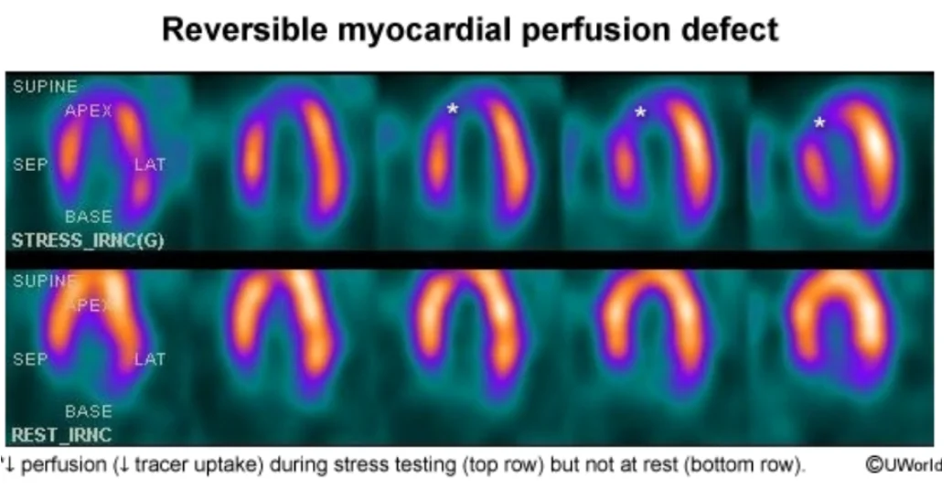
What this shows
Reading the perfusion scan
a technetium-99m tracer (sestamibi or tetrofosmin) enters perfused muscle; rest and stress images are compared.- Normal uptake at rest and stress: no ischemia.
- Reversible defect (reduced with stress, normal at rest): inducible ischemia.
- Fixed defect (reduced at both): scar.
High-risk stress features go to angiography
ST depression at low workload, a fall in systolic blood pressure, poor capacity, ventricular arrhythmia.Management
During an attack
sublingual nitroglycerin is first line.Long term, symptom control
beta blocker is first line.- If beta blockers are contraindicated: a non-dihydropyridine calcium-channel blocker (CCB) or a long-acting nitrate.
- Add a CCB or long-acting nitrate if symptoms persist; ranolazine for refractory angina.
Prevent progression
aspirin and a high-intensity statin for every patient with atherosclerotic cardiovascular disease (ASCVD); both reduce mortality.- Control hypertension and diabetes.
- Add an angiotensin-converting enzyme (ACE) inhibitor or angiotensin receptor blocker (ARB) with hypertension, diabetes, CKD or a left ventricular ejection fraction (LVEF) of 40 percent or less.
Lifestyle
smoking cessation is the most important step, then regular moderate exercise, weight loss and a healthy diet.- The Mediterranean diet (fresh fruit, vegetables, legumes) reduces cardiovascular morbidity and mortality with or without established CAD.
Revascularization
indicated when function stays impaired despite medical therapy, or for high-risk anatomy.- Percutaneous coronary intervention (PCI) is preferred unless coronary artery bypass grafting (CABG) is indicated.
- After a stent, dual antiplatelet therapy (aspirin plus a P2Y12 inhibitor) for 6 to 12 months, then aspirin for life.
CABG indications
three anatomies.- Three-vessel disease: left anterior descending (LAD), right coronary artery (RCA) and left circumflex (LCx).
- Left main stem disease.
- Two-vessel disease involving the LAD in a patient with diabetes.
A vasodilator perfusion scan shows reduced tracer uptake in the anterior wall during stress that is normal at rest. What does that mean, and what should the patient have avoided before the test?
A reversible defect means inducible ischemia (a fixed defect would mean scar). Before a vasodilator test the patient should hold caffeine and other methylxanthines for 12 hours and dipyridamole for 48 hours, as well as beta blockers, calcium-channel blockers and nitrates.
How it's tested
A 55-year-old man with typical exertional angina, a normal resting ECG, and able to walk on a treadmill: exercise ECG stress test.
Same man with a baseline LBBB: vasodilator perfusion imaging or stress echocardiography; the exercise ECG is uninterpretable.
A 58-year-old with claudication and no cardiac symptoms: peripheral artery disease is a CAD equivalent; aspirin, a high-intensity statin and smoking cessation now.
Stable angina in a patient with asthma and a heart rate of 52: amlodipine or a long-acting nitrate; a beta blocker and verapamil or diltiazem are both wrong.
Angina persists on a beta blocker and a nitrate; what improves survival: aspirin and the statin already do; revascularization improves survival only for left main, three-vessel or diabetic multivessel disease.
Diabetic with three-vessel disease and an EF of 35 percent: CABG, not multivessel PCI.
A man on sildenafil takes nitroglycerin for chest pain: profound hypotension; the classic contraindication.
Go deeper
Guidelines: 2023 AHA/ACC Chronic Coronary Disease Guideline
Related Step 2 pages: Acute Coronary Syndrome, Vasospastic Angina, Hyperlipidemia, Coronary Artery Bypass Grafting, Approach to Chest Pain, Approach to Stress Testing
High-yield images2
Flashcards for this page
Card 1 of 5 · try-out only, nothing is saved
You just read one page of 236
The rest of Step 2 CK, written exactly like this.
Rapid Review is the reading layer of a full Step 1 platform: a schedule fitted to your exam date, flashcards for every page, and a mentor a message away.
Every Step 1 system
Written the same way: what to know, how it’s tested, where to go deeper.
A plan built to your exam date
Tell us when you sit, and the schedule fits the whole library and your question bank to it.
Flashcards for every page
Ready-made spaced-repetition decks linked to each topic, so nothing you read gets forgotten.
