
Rapid Review·Cardiovascular
Select any text to highlight it or add a note.
Vascular: Venous
T2High yieldSuperficial Thrombophlebitis
Focus on
A tender, red, palpable cord along a superficial vein — after an IV cannula, in a varicose vein, or migrating in a patient with pancreatic cancer. Usually NSAIDs, warmth and compression; anticoagulate when the clot is long (≥ 5 cm) or near the saphenofemoral junction; and when there is fever and pus at the site it is suppurative thrombophlebitis — pull the line, culture, antibiotics.
Key takeaways
What it is and who gets it
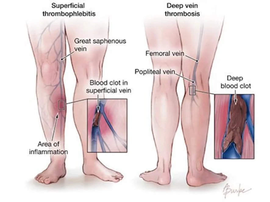
What this shows
Superficial thrombophlebitis
inflammation and thrombosis of a superficial vein, most often in the leg (the great saphenous vein and tributaries) or an arm vein at a cannula site.Lower risk, not no risk
the superficial version of deep vein thrombosis (DVT), with a much lower risk of pulmonary embolism.- But concomitant DVT or embolism is present in about 25 percent, by shared risk factors or, rarely, extension into the deep veins.
Risk factors
the DVT risk factors, varicose veins, venous cannulation and intravenous drugs (an ordinary peripheral cannula is enough), Behcet disease, thromboangiitis obliterans.- A peripherally inserted central catheter causes upper-limb DVT instead, a different disease.
Thrombophlebitis migrans (Trousseau syndrome)
migratory thrombophlebitis of malignancy, classically pancreatic cancer.Clinical features and diagnosis
The cord
pain, tenderness, induration and erythema along a superficial vein with a palpable cord (the thrombosed vein).Little swelling
minimal limb swelling; marked swelling suggests DVT.Diagnosis
clinical.Compression ultrasound
when the diagnosis is unclear or any DVT risk factor is present.- A thickened, edematous, non-compressible superficial vein, and it excludes deep venous involvement.
Migratory disease without varicosities
a cancer evaluation.| Superficial thrombophlebitis | Deep vein thrombosis | Cellulitis | Suppurative thrombophlebitis | |
|---|---|---|---|---|
| Finding | Tender cord along a superficial vein, linear erythema | Diffuse calf or thigh swelling, warmth, tenderness; no cord | Spreading erythema, warmth, edema; no cord; a portal of entry | Cord plus fever, rigors, pus at the cannula site |
| Limb swelling | Minimal | Marked | Moderate, diffuse | Variable |
| Systemic | Low-grade fever at most | None, or embolism symptoms | Fever, leukocytosis | High fever, bacteremia, septic emboli |
| Test | Clinical, ultrasound if a risk factor | Compression ultrasound | Clinical | Blood cultures, ultrasound or CT |
| Treatment | Analgesics, compression; anticoagulate if 5 cm or more or near the deep system | Anticoagulation | Antibiotics | Remove the catheter, intravenous antibiotics, excise the vein if needed |
Management
Symptomatic care
oral or topical analgesics, compression, keep walking, remove the offending cannula.Generally no anticoagulation
it rarely embolizes.- Consider anticoagulation for a thrombus of 5 cm or more, proximity to the deep venous system (within 3 cm of the saphenofemoral junction), or DVT risk factors (prophylactic-dose fondaparinux or low-molecular-weight heparin for 45 days).
Extension into the deep system
treated as DVT.No antibiotics without infection
treat the varicosities after a recurrent episode (Chronic Venous Insufficiency).Suppurative thrombophlebitis and complications
Suppurative thrombophlebitis
venous thrombosis with infection, from an intravascular catheter, injection drug use, or local spread (Lemierre syndrome of the internal jugular after pharyngitis).Features
fever and bacteremia, an erythematous indurated vein, purulent drainage at the catheter site.Diagnosis
blood cultures on removal of the catheter; ultrasound (best initial imaging) or contrast computed tomography (CT) to confirm the thrombus.Treatment
source control (remove the catheter; surgical excision of the vein if needed) and intravenous antibiotics adjusted to sensitivities (Catheter-Related Bloodstream Infection).Complications
extension to DVT and pulmonary embolism; for the suppurative form, septic emboli and persistent bacteremia or fungemia.A woman with varicose veins has a tender, red, 8 cm cord along the great saphenous vein, ending 2 cm from the saphenofemoral junction, with little leg swelling and no fever. What imaging is needed, and does she need anticoagulation?
Superficial thrombophlebitis. Get a compression ultrasound (DVT coexists in about 25 percent). Yes, anticoagulate: the thrombus is 5 cm or more and within 3 cm of the saphenofemoral junction (prophylactic-dose fondaparinux or low-molecular-weight heparin for 45 days).
How it's tested
A woman with varicose veins has a tender red cord on the medial thigh; duplex shows a 7-cm thrombus in the great saphenous vein 2 cm from the saphenofemoral junction: superficial thrombophlebitis with high-risk features — prophylactic fondaparinux (or LMWH) for 45 days, compression, NSAIDs.
A 3-cm tender cord in a calf varicosity, no swelling, no risk factors: NSAIDs, warmth and compression — no anticoagulation.
Recurrent superficial thrombophlebitis at different sites in a 60-year-old smoker with weight loss: Trousseau syndrome — look for pancreatic (or other) adenocarcinoma.
Fever, rigors and pus draining around a peripheral IV placed four days ago, with a tender cord up the forearm: suppurative thrombophlebitis — remove the cannula, blood cultures, IV vancomycin; excise the vein if it does not settle.
Sore throat a week ago, now fever, neck pain and swelling along the sternocleidomastoid with septic lung nodules: Lemierre syndrome — internal jugular septic thrombophlebitis with Fusobacterium.
Go deeper
Guidelines: [2021 CHEST Antithrombotic Therapy for VTE Guideline](https://journal.chestnet.org/article/S0012-3692(21)01506-3/fulltext)
Related Step 2 pages: Chronic Venous Insufficiency, Catheter-Related Bloodstream Infection, Approach to Leg Pain & Ulcers, Approach to Fluid Replacement
High-yield images1
You just read one page of 236
The rest of Step 2 CK, written exactly like this.
Rapid Review is the reading layer of a full Step 1 platform: a schedule fitted to your exam date, flashcards for every page, and a mentor a message away.
Every Step 1 system
Written the same way: what to know, how it’s tested, where to go deeper.
A plan built to your exam date
Tell us when you sit, and the schedule fits the whole library and your question bank to it.
Flashcards for every page
Ready-made spaced-repetition decks linked to each topic, so nothing you read gets forgotten.
