
Rapid Review·Cardiovascular
Select any text to highlight it or add a note.
Vascular: Venous
T2High yieldChronic Venous Insufficiency
Focus on
Heavy aching legs that swell through the day and improve overnight, varicose veins, brown hemosiderin staining and stasis dermatitis at the medial ankle, and eventually a shallow weeping ulcer in the gaiter area. Duplex shows reflux; compression and elevation treat it; ablate the refluxing saphenous vein when conservative care fails. Half of DVTs end here — the post-thrombotic syndrome.
Key takeaways
Pathophysiology and risk factors
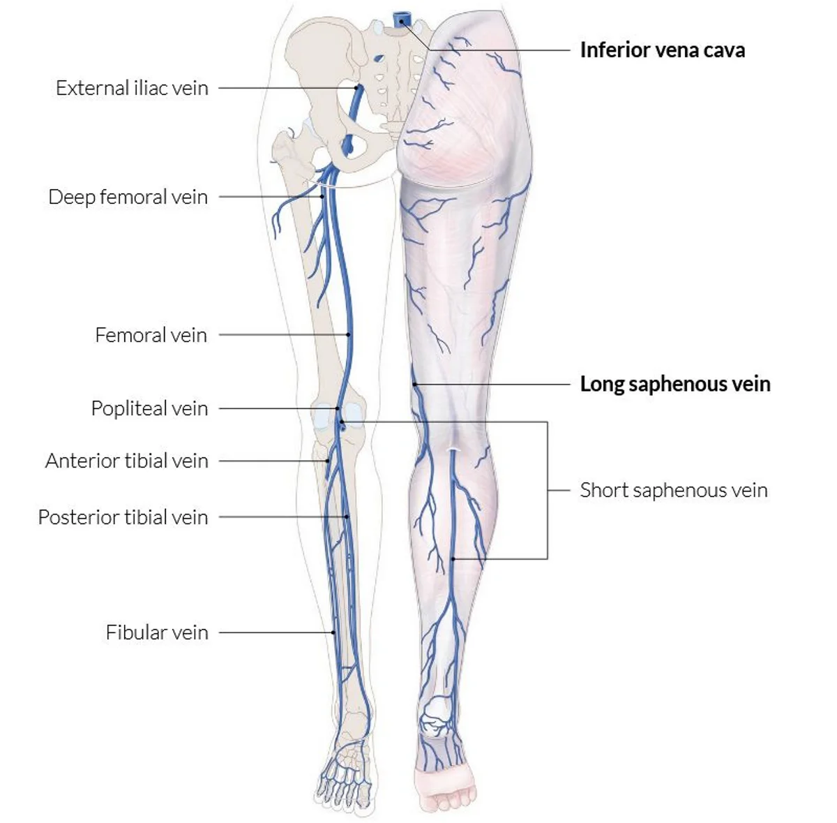
What this shows
Normal flow
runs one way, from the superficial to the deep system through perforating veins, driven by the calf-muscle pump.What goes wrong
elevated venous pressure causes valve incompetence; reflux into the superficial veins raises the pressure further and forms varicose veins; capillary damage then leaks fluid, protein and red cells into the skin.The hallmark
bidirectional flow in perforating veins.Risk factors
advancing age, female sex, smoking, family history, pregnancy, obesity and a sedentary lifestyle, prolonged standing (soldiers), prior limb trauma, and prior deep vein thrombosis (DVT) (the post-thrombotic syndrome).Clinical features and diagnosis
Symptoms
aching, cramping, heaviness and swelling, worse with heat and standing, relieved by walking and leg elevation; pruritus, tingling, numbness.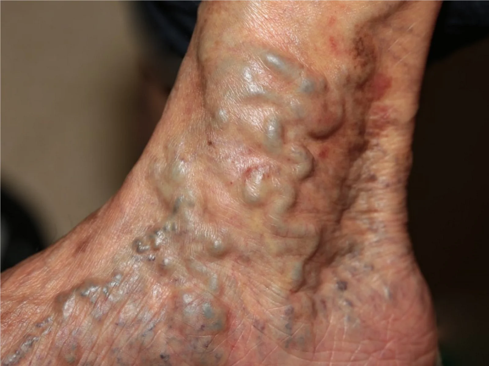
What this shows
Signs
varicose veins (dilated tortuous veins of 3 mm or more), telangiectasias, non-pitting edema starting at the ankle (relative sparing of the feet), yellow-brown or red-brown pigmentation at the medial ankle.Skin changes
stasis dermatitis (the scaly pruritic rash on the Stasis dermatitis) and lipodermatosclerosis (chronic fibrosis of the lower leg).- Skin changes such as dermatitis or an ulcer mean advanced disease.
Local, not systemic
no jugular distension, no crackles; the edema is local.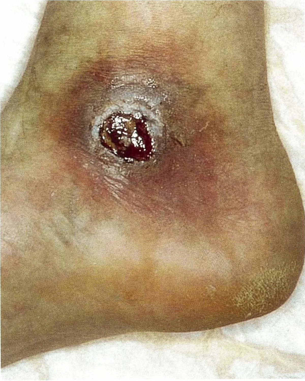
What this shows
Complication
chronic venous stasis ulcers, shallow and exudative in the medial gaiter area.Diagnosis
clinical, from symptoms and examination.Duplex ultrasonography
confirms and stages; bidirectional flow in the great saphenous vein is diagnostic, and it excludes acute thrombosis in a suddenly swollen leg.| Venous insufficiency | Lymphedema | Heart failure or drug edema | Arterial ulcer (Peripheral Artery Disease) | |
|---|---|---|---|---|
| Laterality | Unilateral or bilateral | Usually unilateral | Bilateral | Either |
| Edema | Pitting, then non-pitting; spares the forefoot | Non-pitting, involves the dorsum of the foot and toes | Pitting, symmetric; jugular distension, crackles, or an amlodipine prescription | Absent |
| Skin | Hemosiderin, stasis dermatitis, lipodermatosclerosis, varicosities | Thickened, peau d'orange | Normal | Pale, shiny, hairless, cool |
| Ulcer | Medial gaiter, shallow, exudative, mildly painful | Rare | None | Toes or lateral malleolus, punched-out, dry, painful |
| Key treatment | Compression, elevation, ablation | Decongestive therapy, compression | Diuretics, stop the drug | Revascularization |
Edema by laterality
typically unilateral with DVT, muscle strain, lymphedema, a paralyzed limb or cellulitis; typically bilateral with heart failure, venous insufficiency or a drug (amlodipine).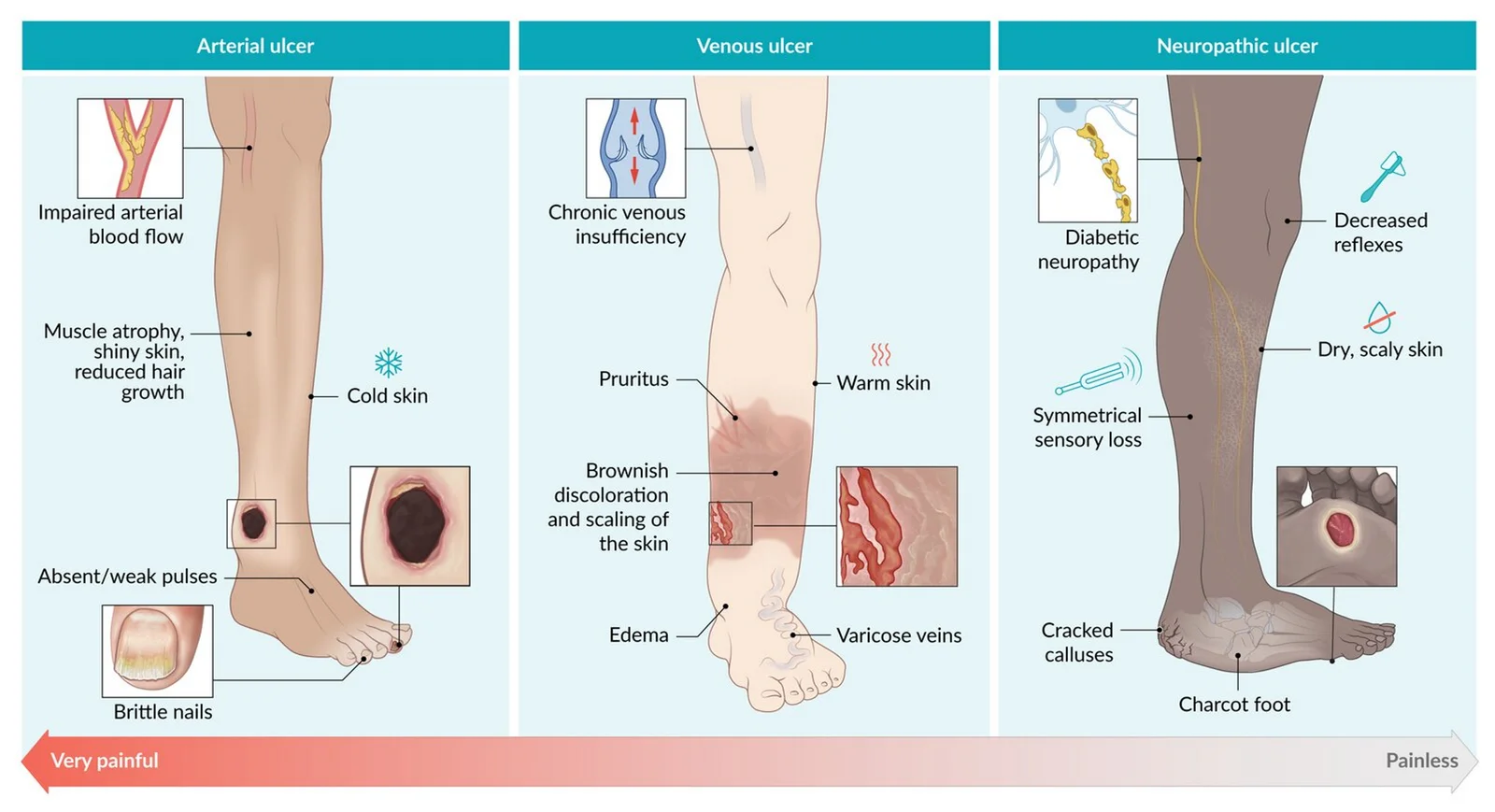
What this shows
Three leg ulcers
arterial, venous and neuropathic.| Arterial ulcer | Venous ulcer | Neuropathic ulcer | |
|---|---|---|---|
| Location | Pressure points of the foot and shin (lateral malleolus, tips of the toes) | Gaiter region above the ankle, usually medial | Plantar pressure points (heel, metatarsal heads) |
| Mechanism | Vessel occlusion and ischemia | Chronic local venous hypertension | Diabetic microvasculopathy and neuropathy |
| Wound | Punched-out, dry | Irregular borders, exudative, superficial | Hyperkeratotic borders, deep |
| Pain | Severe | Mild | Absent |
| Other | Pale, shiny, cold, hairless skin; nail dystrophy; absent pulses | Varicose veins, edema, stasis dermatitis | Charcot joints, absent ankle reflex, lost vibration sense, claw toes |
Management
Conservative measures first
compression stockings, frequent leg elevation, exercise (walking, ankle flexion) to work the calf pump, and avoiding heat and prolonged standing or sitting with bent legs.Stasis dermatitis
emollients and topical steroids, not antibiotics (it is not cellulitis without fever and spreading warmth).Venous ulcers
compression is the treatment, with moist wound care and debridement; pentoxifylline speeds healing; ablate the refluxing veins to prevent recurrence.Refractory cases
endovenous ablation (radiofrequency or laser) of the saphenous vein, sclerotherapy for smaller varicosities, phlebectomy or other surgery.- Ablation needs a patent deep system.
Post-thrombotic syndrome and complications
Post-thrombotic syndrome
chronic venous insufficiency after an acute DVT, in about half of patients within 2 years, from venous hypertension distal to the old thrombus.- Edema, fatigue, pain, superficial venous dilation and ulcers, worse at the end of the day.
- New or worsening swelling needs a venous ultrasound to rule out recurrent DVT.
- Treatment: exercise and compression, the mainstays for venous return.
Complications
chronic recurrent ulcers, stasis and contact dermatitis, cellulitis, Superficial Thrombophlebitis, bleeding from varicosities, DVT.A 58-year-old teacher who stands all day has aching, swollen ankles worse by evening, brown pigmentation at the medial ankles and a shallow weeping ulcer above the medial malleolus; pulses are normal. What confirms the diagnosis, what must be checked before treatment, and what is the treatment?
Chronic venous insufficiency with a venous ulcer. Duplex ultrasound (bidirectional saphenous flow) confirms it. Check the ankle-brachial index before compression; then compression, leg elevation and exercise, moist wound care and pentoxifylline, with ablation for refractory disease.
How it's tested
A 58-year-old woman who stands all day has aching heavy legs that swell by evening, varicose veins, and brown pigmentation with an itchy scaly rash above the medial ankle: chronic venous insufficiency with stasis dermatitis — compression stockings, elevation, exercise, topical steroid; duplex for reflux.
A shallow weeping ulcer above the medial malleolus with surrounding pigmentation and edema, palpable pedal pulses: venous stasis ulcer — check the ABI, then compression bandaging and wound care; pentoxifylline; ablate the refluxing vein.
Unilateral leg swelling and heaviness two years after a femoral DVT, duplex without new thrombus: post-thrombotic syndrome — compression and exercise.
Bilateral non-pitting swelling involving the dorsum of both feet with thickened skin: lymphedema, not venous disease.
Should the red, scaly, weeping venous leg get antibiotics: no — stasis dermatitis is not cellulitis; topical steroids and compression.
Go deeper
Guidelines: [2022 SVS/AVF/AVLS Varicose Vein Guideline](https://www.jvsvenous.org/article/S2213-333X(22)00417-1/fulltext)
Related Step 2 pages: Approach to Leg Pain & Ulcers, Superficial Thrombophlebitis, Peripheral Artery Disease, Heart Failure, Angiosarcoma
High-yield images5
Flashcards for this page
Card 1 of 5 · try-out only, nothing is saved
You just read one page of 236
The rest of Step 2 CK, written exactly like this.
Rapid Review is the reading layer of a full Step 1 platform: a schedule fitted to your exam date, flashcards for every page, and a mentor a message away.
Every Step 1 system
Written the same way: what to know, how it’s tested, where to go deeper.
A plan built to your exam date
Tell us when you sit, and the schedule fits the whole library and your question bank to it.
Flashcards for every page
Ready-made spaced-repetition decks linked to each topic, so nothing you read gets forgotten.
