
Rapid Review·Cardiovascular
Select any text to highlight it or add a note.
Congenital Heart Disease
T1Must knowTruncus Arteriosus
Focus on
One great vessel leaves the heart over a VSD and feeds the aorta, the pulmonary arteries and the coronaries together. Mild cyanosis plus early heart failure from pulmonary flooding, bounding pulses, a single S2, a harsh murmur — and DiGeorge syndrome in a third. Surgical repair in the first weeks before pulmonary vascular disease sets in.
Key takeaways
What it is
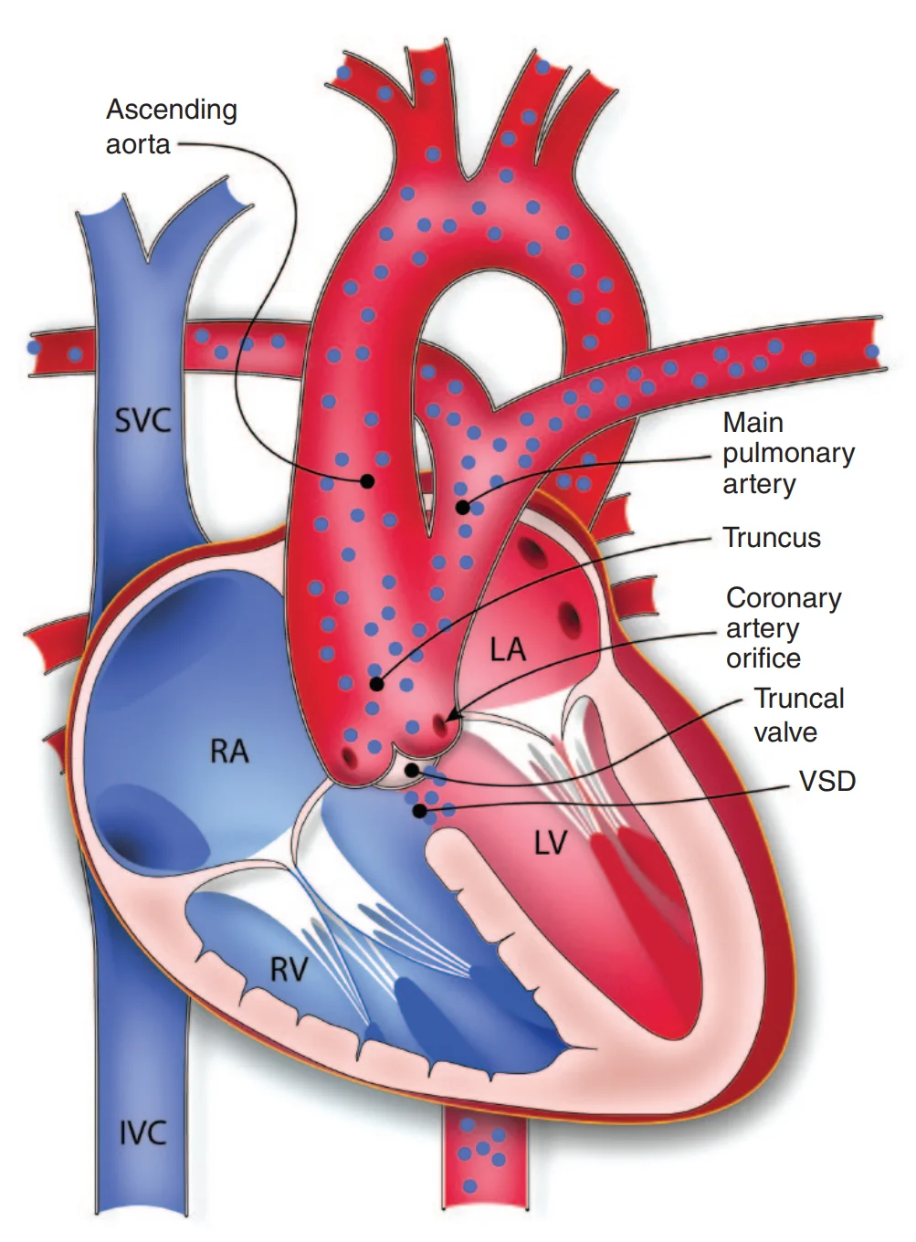
What this shows
Truncus arteriosus
underdevelopment of the aorticopulmonary septum, so the truncus never divides (Cardiac Embryology Essentials).- A single trunk receives the output of both ventricles over a ventricular septal defect (always present) and feeds the coronaries, pulmonary arteries and aorta through one truncal valve.
The physiology
aortic pressure floods the pulmonary arteries (over-circulation and pulmonary edema) and the blood is fully mixed (cyanosis).Associations
DiGeorge syndrome (22q11); maternal diabetes.Clinical features and diagnosis
Cyanosis and respiratory distress in the first weeks of life
from pulmonary edema, then heart failure with failure to thrive as pulmonary resistance falls.Bounding peripheral pulses and a wide pulse pressure
run-off from the trunk into the low-resistance pulmonary bed.The murmur
a harsh systolic murmur at the lower left sternal border with a thrill and a loud single S2 (one valve), often with an ejection click.Transthoracic echocardiography is confirmatory
a single overriding great vessel arising from the heart, the origin of the pulmonary arteries, truncal valve function.Chest film
a right-sided aortic arch, increased pulmonary vascular markings, and an absent thymus if DiGeorge.Electrocardiogram
biventricular hypertrophy.Every case
check calcium and test for the 22q11 deletion (hypocalcemia, T-cell deficiency).| Truncus arteriosus | Transposition of the Great Arteries | Tetralogy of Fallot | |
|---|---|---|---|
| Defect | Single trunk, no aorticopulmonary septum, ventricular septal defect | Great arteries switched | Infundibular stenosis, septal defect, overriding aorta, right ventricular hypertrophy |
| Cyanosis | Mild; heart failure dominates within weeks | Severe within hours | Variable; spells at months |
| Pulses | Bounding, wide pulse pressure | Normal | Normal |
| S2 | Single and loud, with a click | Single and loud | Single (soft P2) |
| Pulmonary markings | Increased | Increased | Decreased |
| Association | DiGeorge, right arch | Maternal diabetes | DiGeorge, Down |
Management and complications
Medical stabilization
diuretics and afterload reduction for the heart failure; correct hypocalcemia.Urgent surgical repair in the first weeks
separate the pulmonary arteries from the trunk, close the defect, and connect the right ventricle to the pulmonary arteries with a valved conduit.Do not delay
delay allows irreversible pulmonary vascular disease (Eisenmenger Syndrome); untreated, most die in infancy.Complications after repair
the conduit does not grow and calcifies, so multiple conduit replacements follow; truncal (neo-aortic) valve regurgitation; branch pulmonary stenosis; arrhythmia; endocarditis.- DiGeorge-related hypocalcemia and infections.
A 3-week-old with mild cyanosis has tachypnea, bounding pulses, a harsh systolic murmur with a click and a single loud S2; the film shows plethoric lungs, a right-sided arch and no thymic shadow. What is the lesion, which electrolyte must be checked, and what is the treatment?
Truncus arteriosus with DiGeorge syndrome (right arch, absent thymus). Check calcium (and test for 22q11). Stabilize the heart failure, then urgent surgical repair in the first weeks.
How it's tested
A 2-week-old with mild cyanosis, tachypnea, sweating with feeds, bounding pulses, a loud single S2 with a click and a harsh systolic murmur; the film shows plethoric lungs and a right aortic arch, and calcium is low: truncus arteriosus with 22q11.2 deletion — echocardiogram, calcium replacement, early complete repair.
Why the bounding pulses and wide pulse pressure: diastolic run-off from the single trunk into the low-resistance pulmonary circulation (plus truncal valve regurgitation).
Which two cyanotic lesions share the DiGeorge association: truncus arteriosus and tetralogy of Fallot (with interrupted aortic arch) — the neural-crest conotruncal family.
Repaired truncus at age 8 with a new harsh systolic murmur and RV hypertension: RV–pulmonary artery conduit stenosis — conduit replacement.
Go deeper
Guidelines: 2018 AHA/ACC Adult Congenital Heart Disease Guideline
Related Step 2 pages: Tetralogy of Fallot, Transposition of the Great Arteries, Approach to Neonatal Cyanosis, Cardiac Embryology Essentials, Approach to Congenital Heart Disease, Eisenmenger Syndrome
High-yield images1
You just read one page of 236
The rest of Step 2 CK, written exactly like this.
Rapid Review is the reading layer of a full Step 1 platform: a schedule fitted to your exam date, flashcards for every page, and a mentor a message away.
Every Step 1 system
Written the same way: what to know, how it’s tested, where to go deeper.
A plan built to your exam date
Tell us when you sit, and the schedule fits the whole library and your question bank to it.
Flashcards for every page
Ready-made spaced-repetition decks linked to each topic, so nothing you read gets forgotten.
