
Rapid Review·Cardiovascular
Select any text to highlight it or add a note.
Congenital Heart Disease
T1Must knowTransposition of the Great Arteries
Focus on
Two parallel circuits: the aorta leaves the right ventricle and the pulmonary artery leaves the left, so oxygenated blood never reaches the body unless there is a hole. Cyanosis within hours of birth, a single loud S2, usually no murmur, an egg on a string on the film — in an infant of a diabetic mother. Prostaglandin E1 and a balloon atrial septostomy to make the circuits mix, then the arterial switch in the first two weeks.
Key takeaways
What it is
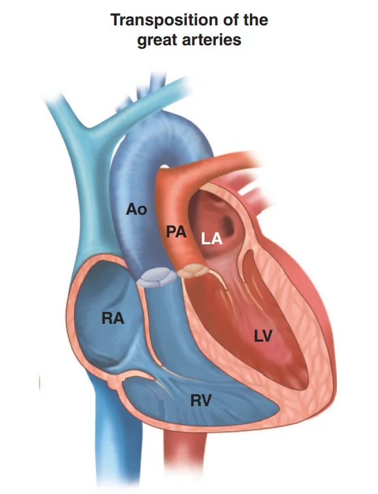
What this shows
D-transposition
the aorta arises from the right ventricle and the pulmonary artery from the left ventricle, because the aorticopulmonary septum failed to spiral (Cardiac Embryology Essentials).Two closed parallel circuits
survival needs mixing through a foramen ovale, ductus or ventricular septal defect (present in about 50 percent, and life-saving).When
the most common cyanotic lesion presenting in the first day of life.Association
maternal diabetes.Clinical features and diagnosis
Cyanosis and respiratory distress within the first 24 hours
worsening as the duct closes; tachypnea, poor feeding.A single loud S2 usually without a murmur
the aorta is anterior, so the pulmonary component is inaudible; a holosystolic murmur only if a ventricular septal defect coexists.Diagnosis
a failed hyperoxia test, then transthoracic echocardiography, which is confirmatory (the great arteries run parallel; the aorta arises anteriorly from the right ventricle).Chest film
"egg on a string" (an egg-shaped heart on a narrow superior mediastinum) with increased pulmonary vascular markings.| D-transposition (complete) | L-transposition (congenitally corrected) | |
|---|---|---|
| Anatomy | Ventricles in normal position; great arteries switched | Ventricles inverted and great arteries switched (two wrongs make a right) |
| Physiology | Parallel circuits; cyanotic, lethal without mixing | Series circulation; acyanotic |
| Presentation | Cyanosis in the first hours of life | Adult failure of the systemic right ventricle, complete heart block, tricuspid regurgitation |
| Treatment | Prostaglandin E1, balloon septostomy, arterial switch | Pacing, heart-failure care, valve surgery |
Management
Allowing the circuits to mix is the life-saving goal
in order.- Alprostadil (prostaglandin E1): maintains the ductus; intubate for apnea if needed.
- Balloon atrial septostomy: creates an artificial atrial septal defect when mixing is inadequate.
- Arterial switch within 2 weeks: reimplanting the coronary arteries, before the left ventricle deconditions against low pulmonary resistance.
Complications
untreated, death from hypoxemia and acidosis in the first weeks.- After the arterial switch: coronary ostial stenosis (the main late concern), supravalvular stenosis at the anastomoses, neo-aortic regurgitation.
The infant of a mother with diabetes is deeply cyanotic 6 hours after birth, with a single loud S2, no murmur, and an egg-shaped heart on a narrow mediastinum. What is the lesion and the first two steps?
D-transposition of the great arteries. First alprostadil (prostaglandin E1) to keep the ductus open, then balloon atrial septostomy if mixing is inadequate; the arterial switch follows within 2 weeks.
How it's tested
A term infant of a poorly controlled diabetic mother is deeply cyanotic at 4 hours; saturation 68% does not improve on 100% oxygen; single loud S2, no murmur; the film shows a narrow mediastinum with an egg-shaped heart: D-transposition — prostaglandin E1 immediately, echocardiogram, balloon atrial septostomy if mixing is inadequate, arterial switch within two weeks.
Why is there no murmur: the great arteries are simply switched — no stenosis or shunt to create turbulence; a murmur appears only with a VSD or pulmonary stenosis.
Why must the arterial switch be done in the first weeks: the left ventricle is pumping against low pulmonary resistance and will deregress; after a few weeks it can no longer support the systemic circulation.
Same infant, slightly less cyanotic, with a holosystolic murmur and hepatomegaly at 3 weeks: TGA with a VSD — better mixing but heart failure from pulmonary over-circulation.
A 35-year-old with congenitally corrected transposition develops complete heart block and heart failure: L-TGA — the systemic morphologic RV failing; pacemaker and heart-failure therapy.
Go deeper
Guidelines: 2018 AHA/ACC Adult Congenital Heart Disease Guideline
Related Step 2 pages: Approach to Neonatal Cyanosis, Approach to Congenital Heart Disease, Fetal Circulation & Transition at Birth, Cardiac Embryology Essentials, Tetralogy of Fallot, Truncus Arteriosus
High-yield images1
You just read one page of 236
The rest of Step 2 CK, written exactly like this.
Rapid Review is the reading layer of a full Step 1 platform: a schedule fitted to your exam date, flashcards for every page, and a mentor a message away.
Every Step 1 system
Written the same way: what to know, how it’s tested, where to go deeper.
A plan built to your exam date
Tell us when you sit, and the schedule fits the whole library and your question bank to it.
Flashcards for every page
Ready-made spaced-repetition decks linked to each topic, so nothing you read gets forgotten.
