
Rapid Review·Cardiovascular
Select any text to highlight it or add a note.
EMBRYOLOGY
T1Must knowAtrial & Ventricular Septation
FA P284-286
Videos
2 to play here
Play 2 here
Focus on
Two septa sliding past each other above, one septum closing below, and the defect each failure leaves behind.
Key takeaways
Atrial septation
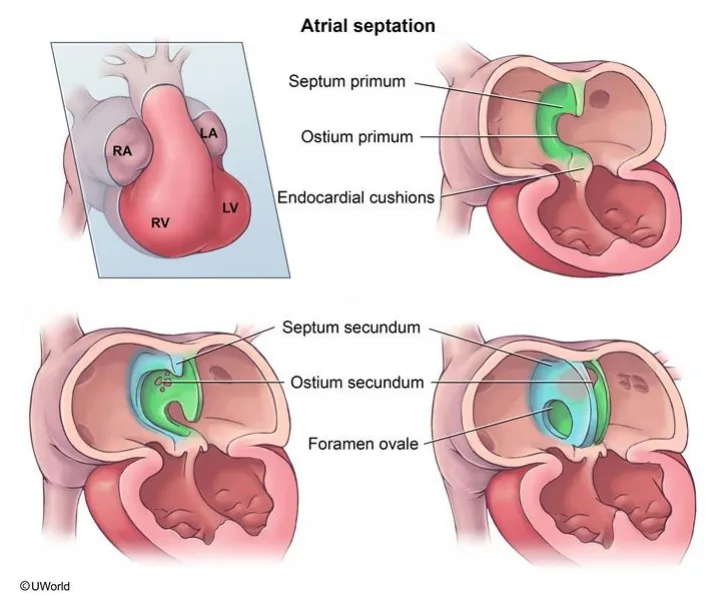
Two septa slide past each other, and the overlap is the foramen ovale
follow the sequence and every atrial defect becomes a named failure of one step.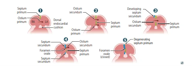
What this shows
What this shows
Septum primum grows toward the endocardial cushions
narrowing the ostium primum.Ostium secundum forms within septum primum by cell death
opening as the ostium primum regresses, so the right-to-left flow is never interrupted.Septum secundum develops to the RIGHT of septum primum
it expands and covers most of the ostium secundum, and the residual opening is the foramen ovale. The remaining portion of septum primum becomes the foramen's one-way valve.At birth the two septa are pressed together
septum primum closes against septum secundum and seals the foramen ovale, because LA pressure rises and RA pressure falls once the lungs inflate. The two fuse during infancy and early childhood to form the definitive atrial septum.PFO is a failure to FUSE, not a hole
present in 25% of the population, and most are asymptomatic and never detected.- It can cause paradoxical emboli, meaning a venous thromboembolus crossing a right-to-left shunt into the systemic circulation and causing stroke, exactly as an ASD can.
- A PFO produces no abnormal heart sound, whereas an ASD gives wide fixed splitting of S2, which is the fastest way to separate them at the bedside.
| Feature | Patent foramen ovale | Atrial septal defect |
|---|---|---|
| Mechanism | Failed fusion of septum primum and secundum | True defect in the interatrial septum |
| Prevalence | 25% of population | Less common |
| Heart sound | None | Wide fixed splitting of S2 |
| Ostium secundum type | Not applicable | Most common, usually isolated |
| Ostium primum type | Not applicable | Rarer; occurs with other anomalies; Down syndrome |
| Shared risk | Paradoxical emboli | Paradoxical emboli |
Ventricular septation
What this shows
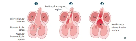
The muscular septum grows UP from the apex
leaving the interventricular foramen open at its top.The aorticopulmonary septum rotates down and fuses with it
forming the membranous interventricular septum and closing the foramen. That junction is the weak point, and it is why the membranous septum is where VSDs occur.Endocardial cushion growth separates atria from ventricles
and contributes to both the atrial septum and the membranous interventricular septum, which is precisely why cushion defects produce combined lesions rather than isolated ones.| Portion | Formed by | Defect |
|---|---|---|
| Muscular interventricular septum | Grows up from the apex | Muscular VSD, most self-resolve |
| Membranous interventricular septum | Aorticopulmonary septum rotates and fuses with muscular septum | Membranous VSD, the most common site |
The defects
Ventricular septal defect is the MOST COMMON congenital cardiac anomaly
usually in the membranous septum. See Congenital Heart Disease: Left-To-Right Shunts.Atrioventricular septal defect is incomplete fusion of the endocardial cushions
also called an endocardial cushion or AV canal defect, giving defects in the septa and in the AV valves.- Complete: a common AV valve with both ASD and VSD.
- Partial: a primum ASD with a cleft mitral valve, causing mitral regurgitation, and no VSD.
- Associated with Down syndrome. Mehlman notes that "endocardial cushion defect" is also used loosely to describe the ASD and VSD seen in Down syndrome.
| Feature | Complete AVSD | Partial AVSD |
|---|---|---|
| Valve | Common AV valve | Cleft mitral valve |
| Septal defects | ASD and VSD | Primum ASD only, no VSD |
| Consequence | Mixing at both levels | Mitral regurgitation |
| Association | Down syndrome | Down syndrome |
A neonate with trisomy 21 has a single large valve orifice between the atria and ventricles on echocardiography, with a defect in the lower interatrial septum and the upper interventricular septum. A second infant with trisomy 21 has a defect in the lower interatrial septum, mitral regurgitation, and an intact ventricular septum. Name each lesion and the single embryologic structure that failed.
Both are atrioventricular septal defects, from failed fusion of the endocardial cushions, and that one structure explains the whole picture. The endocardial cushions build three things: the lower (primum) part of the atrial septum, the membranous part of the interventricular septum, and the AV valves. That is why cushion failure never produces an isolated lesion. The first infant has a complete AVSD, with a common AV valve plus both a primum ASD and a VSD, so there is mixing at the atrial and ventricular levels. The second has a partial AVSD, meaning a primum ASD with a cleft mitral valve producing mitral regurgitation, and crucially no VSD. Note what makes the primum location itself informative: an ostium secundum ASD, the commonest ASD overall, is usually isolated and is a defect of septum primum resorption higher up, whereas a primum defect sits at the cushions and therefore drags valve disease along with it. Both are strongly associated with Down syndrome, and a cleft mitral valve in a trisomy 21 infant is essentially diagnostic of the partial form.
How it's tested
A VSD in the membranous septum is the most common congenital cardiac anomaly, which is the single most likely fact to be asked from this page. For AVSD the association with Down syndrome is the hook, and the partial form is recognised by a cleft mitral valve causing regurgitation with no VSD present. On the atrial side, the exam's favourite point is that a patent foramen ovale is a failure of fusion, not a hole, and that it lets a venous clot reach the systemic circulation as a paradoxical embolus.
Go deeper
First Aid 2026 — CV/Embryology (p.284-286) · B&B — Atrial & ventricular septation · Mehlman — HY Cardio (PFO = ASD on USMLE, paradoxical emboli, Down syndrome AVSD)
High-yield images3
You just read one page of 944
The rest of Step 1, written exactly like this.
Rapid Review is the reading layer of a full Step 1 platform: a schedule fitted to your exam date, flashcards for every page, and a mentor a message away.
Every Step 1 system
Written the same way: what to know, how it’s tested, where to go deeper.
A plan built to your exam date
Tell us when you sit, and the schedule fits the whole library and your question bank to it.
Flashcards for every page
Ready-made spaced-repetition decks linked to each topic, so nothing you read gets forgotten.
