
Rapid Review·Cardiovascular
Select any text to highlight it or add a note.
EMBRYOLOGY
T2High yieldHeart Embryology Derivatives
FA P286
Focus on
What each embryonic structure becomes, and why half of each atrium is smooth and half is trabeculated.
Key takeaways
The organizing idea: smooth versus trabeculated
Smooth walls are absorbed VENOUS inflow; trabeculated walls are the ORIGINAL chamber
deriving this once beats memorizing the table, because the pectinate muscles and the crista terminalis mark exactly where the two meet.- Sinus venosus and pulmonary vein origins give smooth walls.
- Primitive atrium and primitive ventricle origins give trabeculated walls.
- Bulbus cordis gives the smooth outflow tracts.
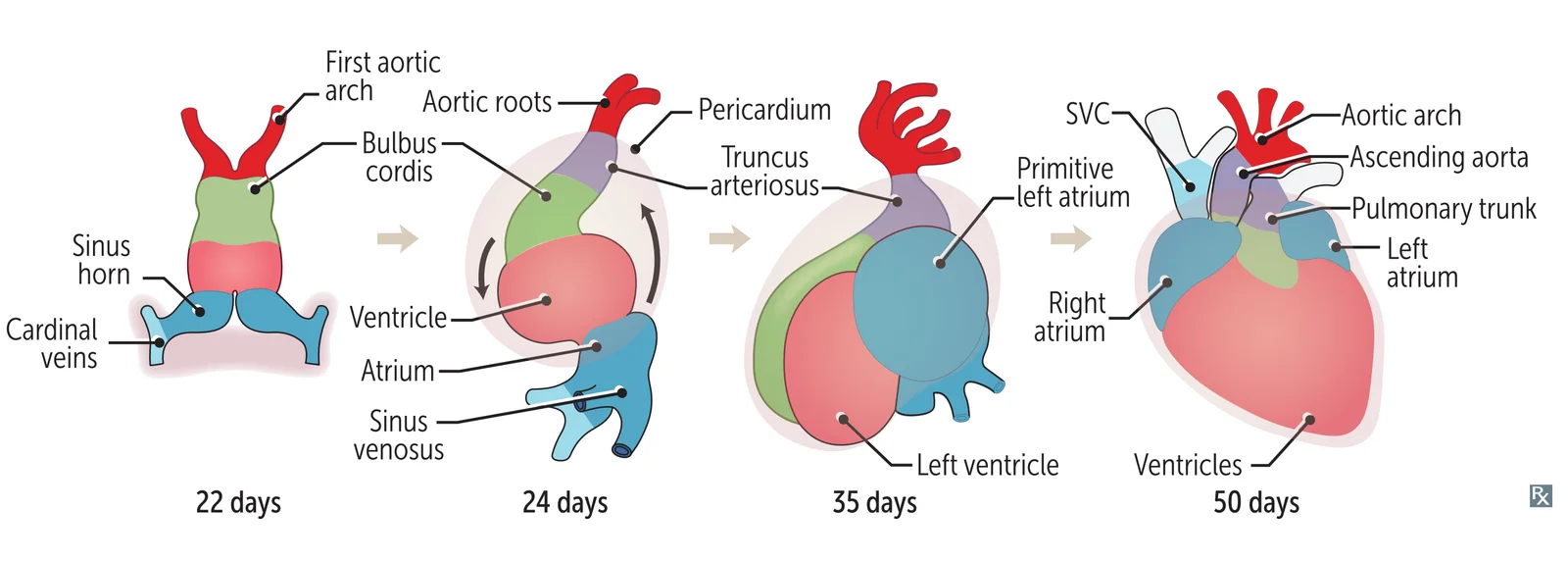
What this shows
The clinical consequence of a failed truncus division
persistent truncus arteriosus, in which the truncus arteriosus fails to divide into the aorta and pulmonary artery before birth. See Heart Tube, Looping & Outflow Tract Formation.| Embryonic structure | Gives rise to |
|---|---|
| Right common / anterior cardinal veins | Superior vena cava |
| Posterior, subcardinal, supracardinal veins | Inferior vena cava |
| Right horn of sinus venosus | Smooth part of RA (sinus venarum) |
| Left horn of sinus venosus | Coronary sinus |
| Primitive pulmonary vein | Smooth part of LA |
| Primitive atrium | Trabeculated parts of LA and RA |
| Endocardial cushion | Atrial septum, membranous IV septum, AV and semilunar valves |
| Primitive ventricle | Trabeculated parts of LV and RV |
| Bulbus cordis | Smooth outflow tracts of LV and RV |
| Truncus arteriosus | Ascending aorta and pulmonary trunk |
Where the great veins come from
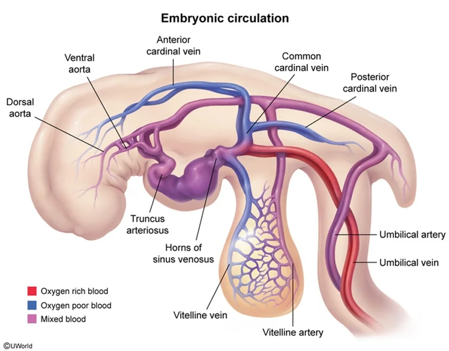
What this shows
Three venous systems drain into the sinus venosus, and each has its own fate
the cardinal veins become the systemic venae cavae, the vitelline veins become the portal system, and the umbilical veins carry placental blood and leave the ligamentum teres.The coronary sinus derives from the LEFT horn of the sinus venosus
it runs in the left AV groove and drains into the right atrium. The right horn is absorbed instead into the smooth-walled sinus venarum of the right atrium.The aortic arches
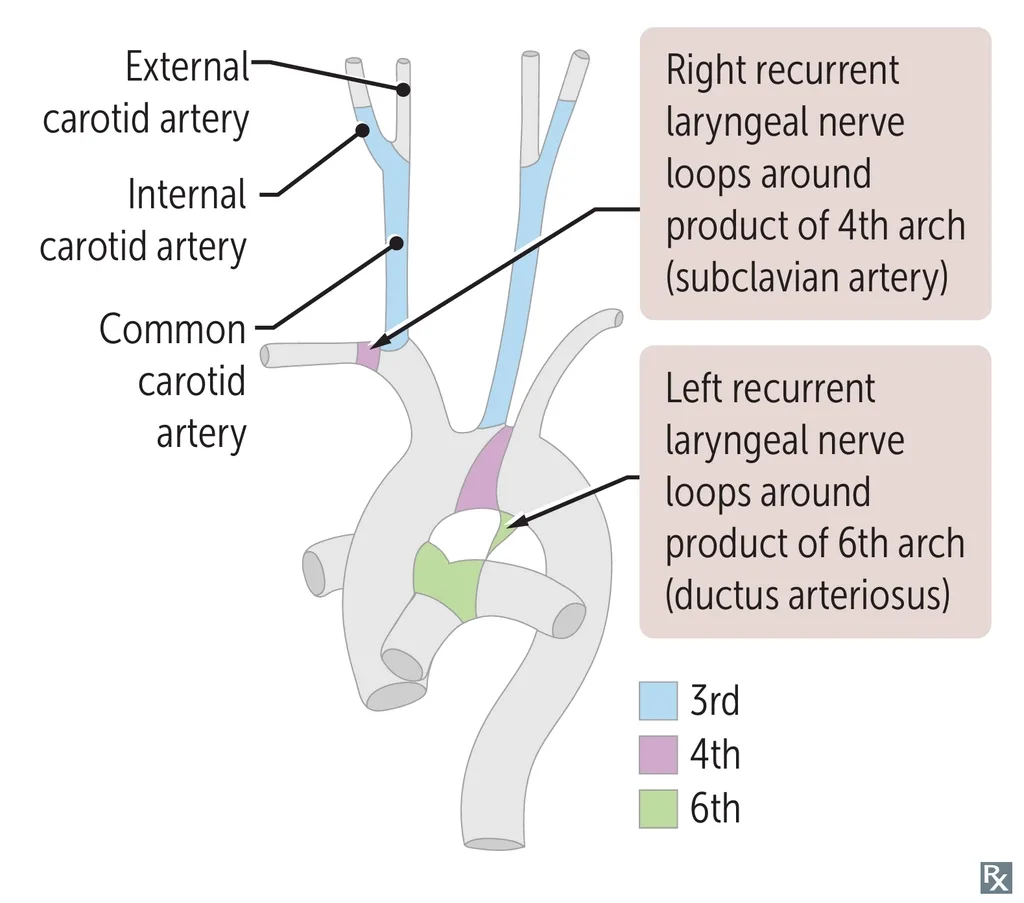
What this shows
The arches are learned by mnemonic, one line each
- 1st arch: part of the maxillary artery, a branch of the external carotid. The 1st arch is maximal.
- 2nd arch: stapedial artery and hyoid artery. Second = Stapedial.
- 3rd arch: common carotid and the proximal internal carotid. C is the 3rd letter.
- 4th arch: left forms the aortic arch, right forms the proximal right subclavian artery. The 4th arch has 4 limbs, so it is systemic.
- 6th arch: right forms the right pulmonary artery, left forms the left pulmonary artery and the ductus arteriosus. The 6th arch is pulmonary, plus the pulmonary-to-systemic shunt.
An echocardiogram in a 60-year-old with long-standing mitral stenosis shows a markedly dilated left atrium. He has become hoarse and has difficulty swallowing solids. Trace both symptoms back to embryology, and say which structure the coronary sinus came from while you are there.
Both symptoms come from the left atrium being the most posterior chamber, pressing on two structures behind it. The dysphagia is direct compression of the esophagus. The hoarseness is compression of the left recurrent laryngeal nerve, and this is Ortner syndrome. The embryology explains why it is the left nerve: each recurrent laryngeal nerve hooks under the last persisting aortic arch derivative on its side, and because the left 6th arch persists as the ductus arteriosus, the left nerve is pulled deep into the thorax to loop under the ligamentum arteriosum, right beside the enlarging atrium. On the right the 6th arch regresses, so that nerve only reaches the 4th arch derivative, the right subclavian artery, and stays safely in the neck. As for the coronary sinus, it derives from the left horn of the sinus venosus, and it too runs posteriorly, in the left atrioventricular groove, which is why a dilated coronary sinus appears on the same echocardiogram whenever right atrial pressure is raised. Contrast that with the right horn of the sinus venosus, which is absorbed to become the smooth-walled sinus venarum of the right atrium, the part separated from the trabeculated original atrium by the crista terminalis.
How it's tested
The derivative table is asked directly, most often for the coronary sinus, which comes from the left horn of the sinus venosus, and for the smooth part of the right atrium, which comes from the right horn. The organizing rule is worth deriving rather than memorising: the smooth-walled portion is always the absorbed venous inflow and the trabeculated portion is always the original primitive chamber, which is why the pectinate muscles and the crista terminalis mark the boundary between them.
Go deeper
First Aid 2026 — CV/Embryology (p.286) · B&B — Cardiac derivatives · Mehlman — HY Cardio (fetal remnants)
High-yield images3
Flashcards for this page
Card 1 of 5 · try-out only, nothing is saved
(...) horn of (...)
You just read one page of 944
The rest of Step 1, written exactly like this.
Rapid Review is the reading layer of a full Step 1 platform: a schedule fitted to your exam date, flashcards for every page, and a mentor a message away.
Every Step 1 system
Written the same way: what to know, how it’s tested, where to go deeper.
A plan built to your exam date
Tell us when you sit, and the schedule fits the whole library and your question bank to it.
Flashcards for every page
Ready-made spaced-repetition decks linked to each topic, so nothing you read gets forgotten.
