
Rapid Review·General Pathology
Select any text to highlight it or add a note.
CELLULAR INJURY
T2High yieldAmyloidosis
P208
Focus on
Any protein that misfolds into a β-pleated sheet, deposits extracellularly, and strangles the organ it lands in.
Key takeaways
One fibril, many proteins
The common mechanism
extracellular deposition of protein in an abnormal fibrillar, β-pleated sheet configuration → cell injury and apoptosis. The precursor protein differs by disease, but the fibril architecture never does.How to see it
Stains
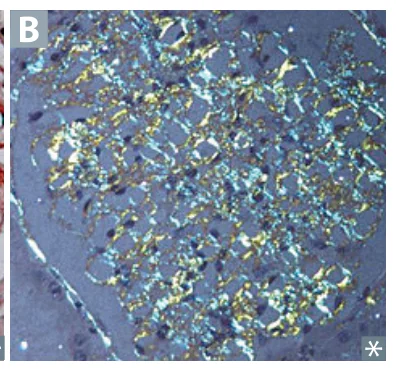
Congo red gives red-orange under normal light and apple-green birefringence under polarized light. Amorphous pink on H&E.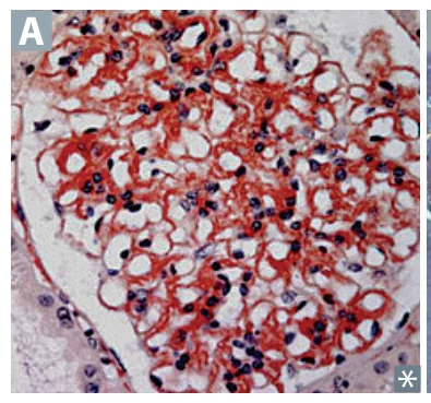
What it does to each organ
Manifestations by organ
- Renal → nephrotic syndrome, the most common presentation.
- Cardiac → restrictive cardiomyopathy.
- GI → hepatosplenomegaly.
- Neuro → dementia, peripheral neuropathy.
- MSK → macroglossia, carpal tunnel syndrome.
- Skin → waxy thickening, easy bruising with periorbital purpura.
Naming the amyloid
| Type | Fibril Protein | Precursor / Association |
|---|---|---|
| AL (Primary) | Ig Light chains | Plasma cell dyscrasias (multiple myeloma) |
| AA (Secondary) | Serum Amyloid A | Chronic inflammation: RA, IBD, familial Mediterranean fever, protracted infection ("A" for inflammation) |
| Transthyretin (ATTR) | Transthyretin | Sporadic (wild-type) in aging, mainly cardiac; hereditary (mutant) causes familial amyloid polyneuropathy/cardiomyopathy |
| Dialysis-related | β2-microglobulin | ESRD on long-term dialysis → carpal tunnel |
| Alzheimer disease | β-amyloid | Cleaved from APP on chromosome 21, hence early onset in Down syndrome |
| Isolated atrial | ANP | Aging → ↑ risk of atrial fibrillation |
| Type 2 diabetes | Islet amyloid polypeptide (amylin) | Deposits in pancreatic islets |
| Medullary thyroid CA | Calcitonin | Secreted by the tumor cells themselves |
A man on hemodialysis for 15 years develops bilateral carpal tunnel syndrome. Which protein makes up the amyloid?
β2-microglobulin, which is not cleared by long-term dialysis in end-stage renal disease and deposits as dialysis-related amyloid.
How it's tested
Systemic amyloidosis is diagnosed by tissue biopsy, classically an abdominal fat pad aspirate, showing Congo red staining with apple-green birefringence under polarized light. That is the answer to "most appropriate next step" whenever a patient has nephrotic-range proteinuria plus a restrictive cardiomyopathy plus macroglossia.
The mechanistic layer: in AA amyloidosis, chronic IL-6-driven hepatic production of serum amyloid A over years is what deposits, so prolonged elevation of an acute-phase reactant is the direct cause. That is why longstanding RA or familial Mediterranean fever is always sitting in the stem.
Go deeper
First Aid "Amyloidosis"; Pathoma Ch. 2 "Amyloidosis"; Boards & Beyond. Anchor the Congo red apple-green birefringence and the source-protein map (AL = light chains/myeloma; AA = SAA/chronic inflammation; β2-microglobulin = dialysis; Aβ = Alzheimer; ATTR = cardiac).
High-yield images2
Flashcards for this page
Card 1 of 5 · try-out only, nothing is saved
(...) fever
(...) disease
(...)
You just read one page of 944
The rest of Step 1, written exactly like this.
Rapid Review is the reading layer of a full Step 1 platform: a schedule fitted to your exam date, flashcards for every page, and a mentor a message away.
Every Step 1 system
Written the same way: what to know, how it’s tested, where to go deeper.
A plan built to your exam date
Tell us when you sit, and the schedule fits the whole library and your question bank to it.
Flashcards for every page
Ready-made spaced-repetition decks linked to each topic, so nothing you read gets forgotten.
