
Rapid Review·General Pathology
Select any text to highlight it or add a note.
INFLAMMATION
T2High yieldWound Healing & Scar Formation
P212–P214
Focus on
The growth factors and phases that close a wound, and what happens when regeneration is impossible and connective tissue fills the gap.
Key takeaways
Regenerate or scar
- Labile cells (gut epithelium, skin, bone marrow) divide continuously and regenerate fully.
- Stable/quiescent cells (hepatocytes, renal tubules) sit in G0 and re-enter the cycle when stimulated.
- Permanent cells (neurons, cardiac and skeletal muscle) cannot regenerate, so the tissue can only scar.
The four phases
Phases of wound healing, in order
- Hemostasis (immediate): vasoconstriction driven by endothelin, platelet plug, coagulation cascade.
- Inflammatory (up to 3 days): platelets, then neutrophils first, then macrophages clear debris roughly 2 days later. Clot formation and ↑ vessel permeability.
- Proliferative (day 3 to weeks): fibroblasts, myofibroblasts, endothelial cells, keratinocytes, macrophages. Granulation tissue plus type III collagen, angiogenesis, epithelial proliferation, clot dissolution, and wound contraction by myofibroblasts. Delayed in vitamin C and copper deficiency.
- Remodeling (1 week to 6+ months): fibroblasts replace type III collagen with type I → ↑ tensile strength. Collagenases require zinc, so zinc deficiency delays healing.
The growth factors
| Mediator | Role |
|---|---|
| FGF | Stimulates angiogenesis, fibroblast proliferation, collagen synthesis |
| TGF-β | Angiogenesis plus fibrosis; ↑ fibroblast migration and proliferation, ↑ collagen and fibronectin synthesis, ↓ ECM degradation by metalloproteinases |
| VEGF | Stimulates angiogenesis; induced by local ↓ pO₂ |
| PDGF | Secreted by activated platelets and macrophages; drives vascular remodeling and smooth muscle migration; recruits fibroblasts |
| Metalloproteinases | Tissue remodeling; require zinc |
| EGF | Stimulates cell growth via tyrosine kinases (EGFR/ErbB1); re-epithelialization |
Collagen and tensile strength
The collagen switch is what the tensile-strength curve actually measures
- Type III collagen is laid down early in granulation tissue: pink, weak, and disorganized.
- Type I collagen replaces it during remodeling: white, strong, and the dominant collagen of bone and mature scar.
- Remodeling needs zinc-dependent collagenases; hydroxylation needs vitamin C; cross-linking by lysyl oxidase needs copper.
When scarring happens
repair cannot be accomplished by cell regeneration alone, so nonregenerated cells, lost to severe acute or chronic injury, are replaced by connective tissue.- 70 to 80% of tensile strength is regained at 3 months, and little is gained after that. A scar never reaches 100%.
- Excess TGF-β drives aberrant scarring of both kinds, hypertrophic and keloid.
Hypertrophic scar vs keloid
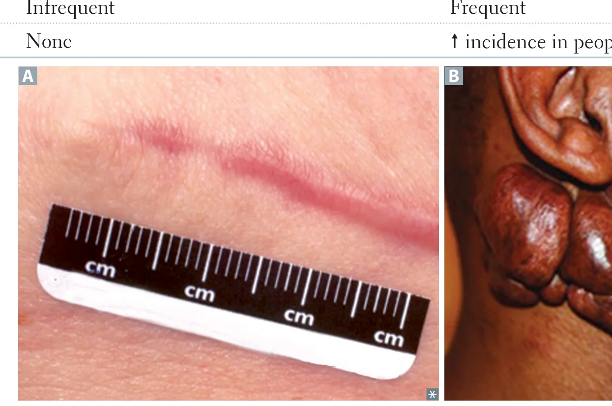
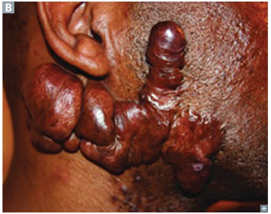
| Feature | Hypertrophic Scar | Keloid Scar |
|---|---|---|
| Collagen synthesis | ↑ (type III > type I) | ↑↑↑ (type I > type III) |
| Collagen organization | Parallel | Disorganized |
| Extent | Confined to the borders of the original wound | Extends BEYOND the borders, with clawlike projections on earlobes, face, upper extremities |
| Recurrence | Infrequent | Frequent |
| Predisposition | None | ↑ incidence in people with darker skin |
Nerve healing
- PNS regenerates because Schwann cells remyelinate; the CNS does not, because oligodendrocytes do not effectively remyelinate after injury.
- The optic nerve is CNS tissue myelinated by oligodendrocytes, which is why optic nerve degeneration causes permanent blindness while a CN VII palsy commonly recovers.
Six weeks after surgery, a wound reopens in a patient with poor nutrition. Which collagen type dominates a wound at this stage, and which deficiencies delay healing?
The wound is still converting type III collagen (granulation tissue) to type I during remodeling. Vitamin C and copper deficiency delay the proliferative phase, and zinc deficiency delays remodeling.
How it's tested
A stem describes a cytokine that increases fibroblast proliferation, increases collagen and fibronectin synthesis, and decreases ECM degradation by metalloproteinases. That triple action is TGF-β, the mediator of both normal fibrosis and pathologic keloid scarring.
If asked which cell initiates fibrosis, the answer is the macrophage. If asked which cell mediates wound contraction, it is the myofibroblast.
The single distinguishing fact is whether the scar respects the original wound margin. A raised scar that stays exactly within the borders of a surgical incision is hypertrophic (type III > type I, parallel bundles). A nodular mass on the earlobe after piercing that has grown well past the puncture with clawlike extensions is a keloid (type I > type III, disorganized, frequently recurs after excision). Both are driven by excess TGF-β.
Go deeper
First Aid "Wound healing" phases + "Scar formation"; Pathoma Ch. 3 "Tissue repair"; Boards & Beyond. Anchor the labile/stable (regenerate) vs permanent (scar only) cell classes, the collagen switch (weak type III laid down early → remodeled to strong type I, needs zinc-dependent collagenase), the cofactors (vitamin C for hydroxylation, copper for lysyl oxidase cross-linking), and "hypertrophic = type III, stays within borders; keloid = excess type I + III, grows BEYOND borders."
High-yield images2
Flashcards for this page
Card 1 of 5 · try-out only, nothing is saved
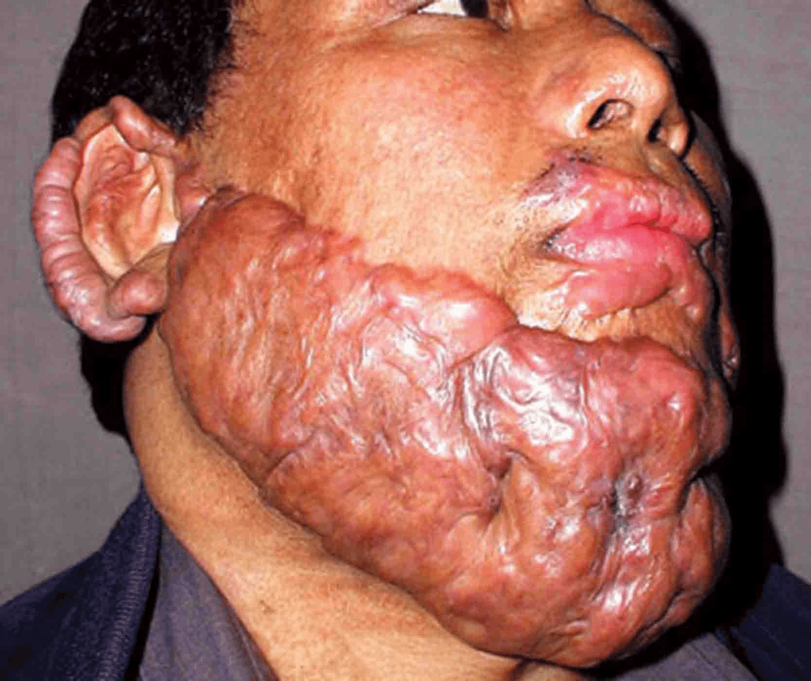
(...)
You just read one page of 944
The rest of Step 1, written exactly like this.
Rapid Review is the reading layer of a full Step 1 platform: a schedule fitted to your exam date, flashcards for every page, and a mentor a message away.
Every Step 1 system
Written the same way: what to know, how it’s tested, where to go deeper.
A plan built to your exam date
Tell us when you sit, and the schedule fits the whole library and your question bank to it.
Flashcards for every page
Ready-made spaced-repetition decks linked to each topic, so nothing you read gets forgotten.
