
Rapid Review·Cardiovascular
Select any text to highlight it or add a note.
Vascular: Peripheral
T1Must knowAcute Limb Ischemia
Focus on
The six Ps — pain, pallor, pulselessness, poikilothermia, paresthesia, paralysis — with sensory loss early and motor loss late. Heparin at once on clinical grounds, then Doppler to grade the threat: a viable limb gets CT angiography and revascularization within 24 hours, a threatened limb goes straight to the operating room within 6 hours, an insensate paralyzed limb is amputated. Embolus (AF) gets embolectomy; thrombosis gets catheter-directed lysis.
Key takeaways
What it is and what causes it
Acute limb ischemia
a sudden, critical fall in limb perfusion; nerve and muscle tolerate only about 6 hours of complete ischemia.Embolism (sudden onset)
a cardiac source in most, above all atrial fibrillation, then mural thrombus after infarction, valve disease, endocarditis, myxoma.- Emboli lodge at bifurcations, the femoropopliteal region most often.
Thrombosis (slower onset)
plaque disruption in Peripheral Artery Disease (acute-on-chronic), or thrombosis of a popliteal or femoral aneurysm.- Because collaterals exist, it presents less dramatically (over 24 hours, prolonged rather than absent capillary refill), but still needs emergency intervention.
Other causes
trauma with vascular injury, and rarely phlegmasia (massive venous thrombosis choking arterial inflow).Acute aortic occlusion
a saddle embolus at the aortic bifurcation (atrial fibrillation, recent infarction) causes bilateral acute lower-limb ischemia.- CT angiography confirms it; treatment is transfemoral embolectomy, aortobifemoral bypass or thrombolysis.
Clinical features
The six P's
pain, pallor, pulselessness, poikilothermia (cold), paresthesia, paralysis.Sensory loss is early; motor loss is late
motor loss means the limb is in jeopardy.Embolic vs thrombotic
embolic is abrupt, with a normal contralateral limb and an irregular pulse; thrombotic has prior claudication, bilateral pulse deficits and a slower course.Diagnosis
Mainly clinical: start heparin immediately
in a classic presentation (the only exception is a contraindication to anticoagulation).Best initial test
arterial and venous Doppler, done after heparin, to grade the threat (the table below).Digital subtraction angiography is the confirmatory test
and the route to intervention; computed tomography (CT) angiography maps a viable limb.Find the source
electrocardiogram and echocardiography (atrial fibrillation, a ventricular thrombus after a recent infarction).| Stage | Sensory loss | Motor weakness | Arterial Doppler | Venous Doppler | Treatment |
|---|---|---|---|---|---|
| I (viable) | None | None | Audible | Audible | CT angiography to define the obstruction, then urgent revascularization within 6 to 24 hours |
| IIb (threatened) | Extends beyond the toes | Mild to moderate | Inaudible | Audible | Emergent revascularization within 6 hours; no time for CT |
| III (non-viable) | Insensate limb | Paralysis | Inaudible | Inaudible | Amputation (revascularizing dead muscle causes lethal reperfusion injury) |
Management
Everyone
heparin, analgesia, fluids, keep the limb dependent and warm (no elevation).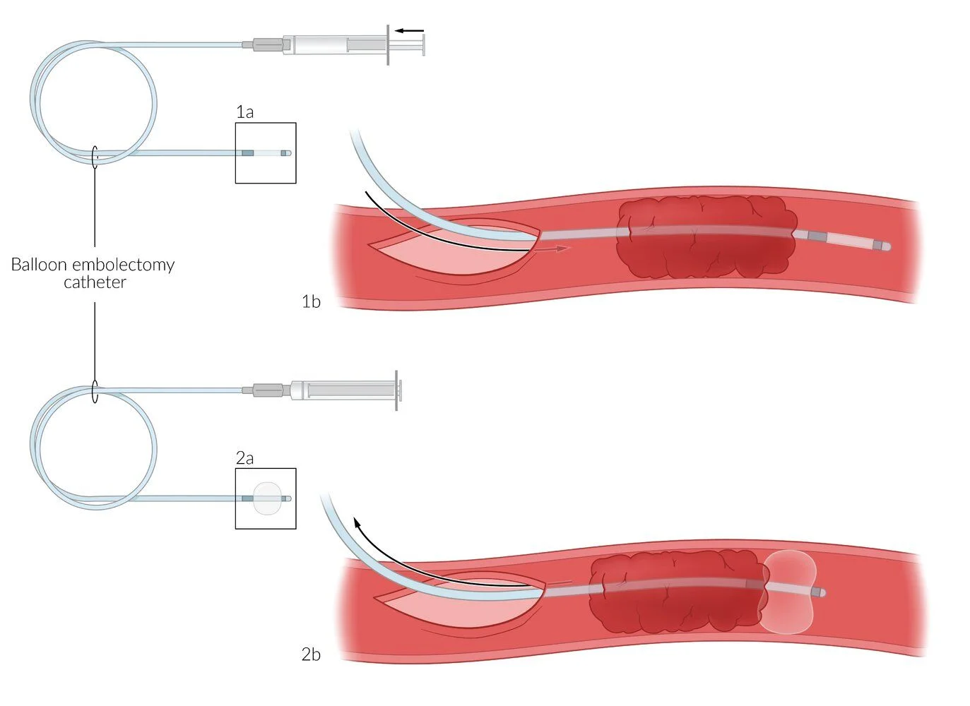
What this shows
Embolic occlusion
surgical balloon-catheter (Fogarty) embolectomy.Thrombotic occlusion
catheter-directed thrombolysis, then treat the underlying stenosis with angioplasty, stent or bypass.- Lysis needs a viable limb with an audible venous signal and no bleeding contraindication.
Gangrene
means amputation.After reperfusion
watch for compartment syndrome (fasciotomy, often prophylactic after 4 to 6 hours of ischemia) and reperfusion injury (rhabdomyolysis, hyperkalemia, myoglobinuric kidney injury, acidosis): fluids and treat the potassium.Long term
anticoagulation for atrial fibrillation or a cardiac thrombus; antiplatelet and statin for peripheral artery disease; repair a popliteal aneurysm.Complications
limb loss, compartment syndrome, reperfusion syndrome with renal failure, recurrent embolism, and death from the cardiac disease that caused the embolus.A 74-year-old with atrial fibrillation has a sudden cold, pale, pulseless right leg with numbness beyond the toes and mild weakness; the arterial Doppler is inaudible but the venous Doppler is audible. What is the first drug, the stage, and the definitive treatment?
Embolic acute limb ischemia, Rutherford IIb (threatened). Start heparin immediately, then emergent revascularization within 6 hours (no time for CT): balloon-catheter embolectomy for an embolus. Watch for compartment syndrome and reperfusion injury afterwards.
How it's tested
A woman with atrial fibrillation not on anticoagulation develops sudden severe pain, pallor and coldness of the left leg with no popliteal pulse; she can still move her toes but feels numb: embolic acute limb ischemia, class IIa–IIb — IV heparin now, then emergency surgical embolectomy; echo for a source afterward.
Same leg, now paralyzed, insensate, mottled and rigid with no venous Doppler signal: irreversible (class III) — amputation, not revascularization.
A man with long-standing claudication whose calf pain became constant over a day, foot cool with prolonged but present capillary refill: acute-on-chronic thrombosis — heparin, angiography, catheter-directed thrombolysis and treatment of the culprit stenosis.
Six hours after successful embolectomy the calf becomes tense and exquisitely painful with paresthesias: compartment syndrome — fasciotomy.
Which is an early sign and which a late sign: sensory loss early, paralysis late.
Go deeper
Guidelines: 2024 ACC/AHA Peripheral Artery Disease Guideline
Related Step 2 pages: Peripheral Artery Disease, Approach to Leg Pain & Ulcers, Atrial Fibrillation, Approach to Aortic Syndromes, Femoral Artery Disease, Vascular Trauma
High-yield images1
Flashcards for this page
Card 1 of 3 · try-out only, nothing is saved
You just read one page of 236
The rest of Step 2 CK, written exactly like this.
Rapid Review is the reading layer of a full Step 1 platform: a schedule fitted to your exam date, flashcards for every page, and a mentor a message away.
Every Step 1 system
Written the same way: what to know, how it’s tested, where to go deeper.
A plan built to your exam date
Tell us when you sit, and the schedule fits the whole library and your question bank to it.
Flashcards for every page
Ready-made spaced-repetition decks linked to each topic, so nothing you read gets forgotten.
