
Rapid Review·Cardiovascular
Select any text to highlight it or add a note.
Vascular: Aorta & Carotid
T1Must knowTraumatic Aortic Injury
Focus on
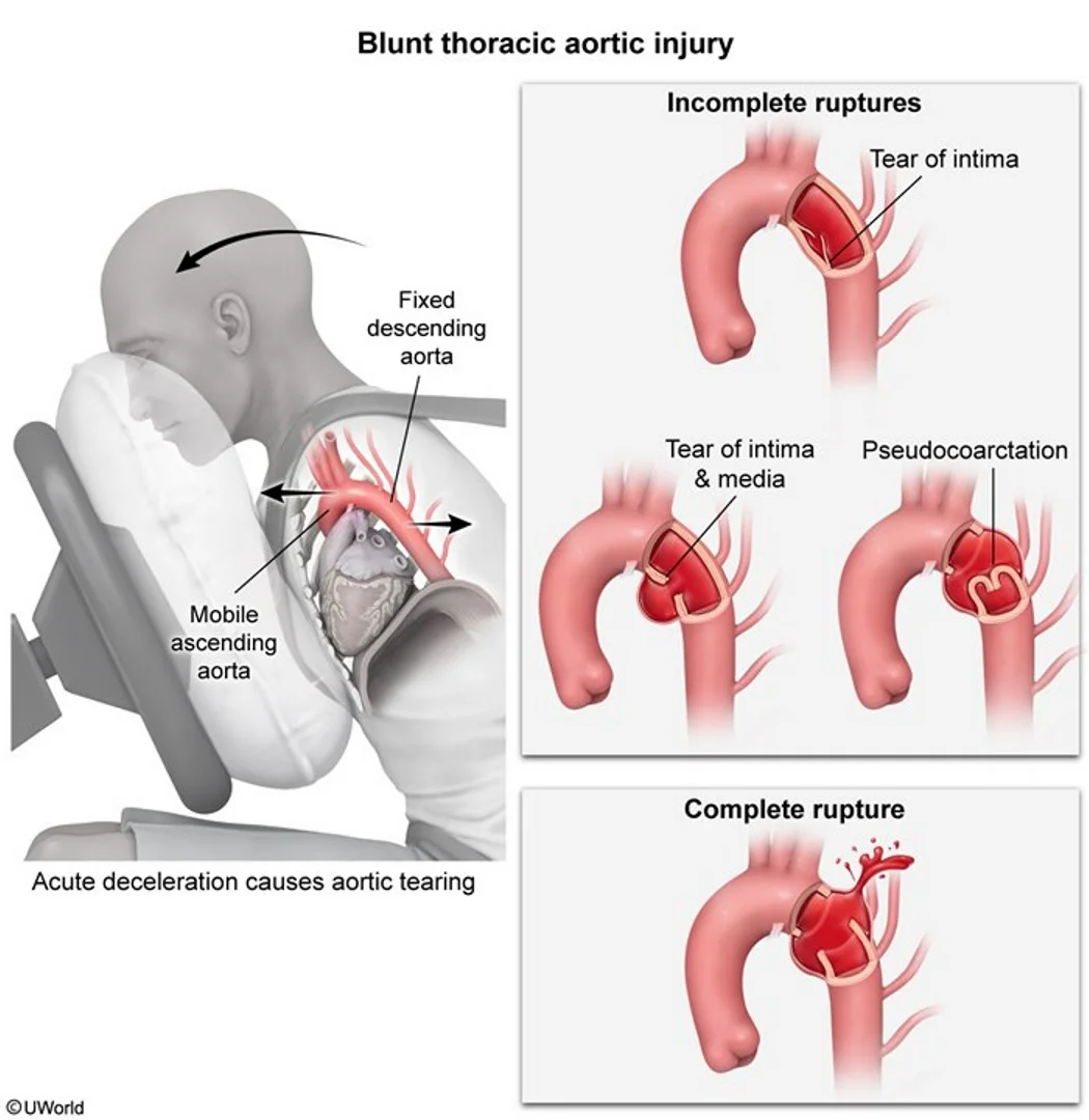
High-energy deceleration — a head-on crash or a fall from over three meters — tears the aorta at the isthmus, just beyond the left subclavian. The patient may look stable. A widened mediastinum on the trauma film mandates CT angiography; keep the pressure and heart rate down and repair it, usually with a stent graft.
Key takeaways
What it is
What this shows
Traumatic aortic injury
a shear tear from high-energy blunt trauma with rapid deceleration (a motor vehicle crash, a fall over 3 m).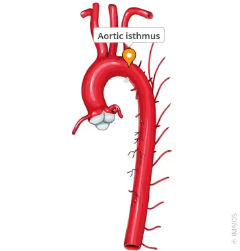
What this shows
Where
most often at the aortic isthmus (proximal descending aorta just distal to the left subclavian origin).Survival
complete transection kills at the scene; survivors have a partial tear contained by the adventitia that can rupture at any moment.Clinical features
It may look stable
an incomplete rupture may appear stable, normotensive or hypertensive from the sympathetic response, despite a life-threatening injury.Clinically overt injury
anxiety, tachycardia and hypotension (hemorrhagic shock), a left hemothorax.Clues
interscapular pain, pulse or pressure asymmetry between the arms, upper-limb hypertension with weak femoral pulses, paraplegia, external chest trauma (seat-belt bruise, sternal or first-rib fractures).Diagnosis
Chest radiograph is the initial screening test
for every suspected blunt aortic injury (deceleration mechanism, fall over 10 feet).- A widened mediastinum is the most sensitive finding; also an abnormal aortic knob, a left effusion from hemothorax, tracheal deviation.
- Compare on the Review the chest radiograph findings.
- A normal film does not exclude injury when the mechanism is severe.
Confirm
computed tomography (CT) angiography in the stable patient, or transesophageal echocardiography (TEE) in the unstable, hypotensive patient (often in the operating room).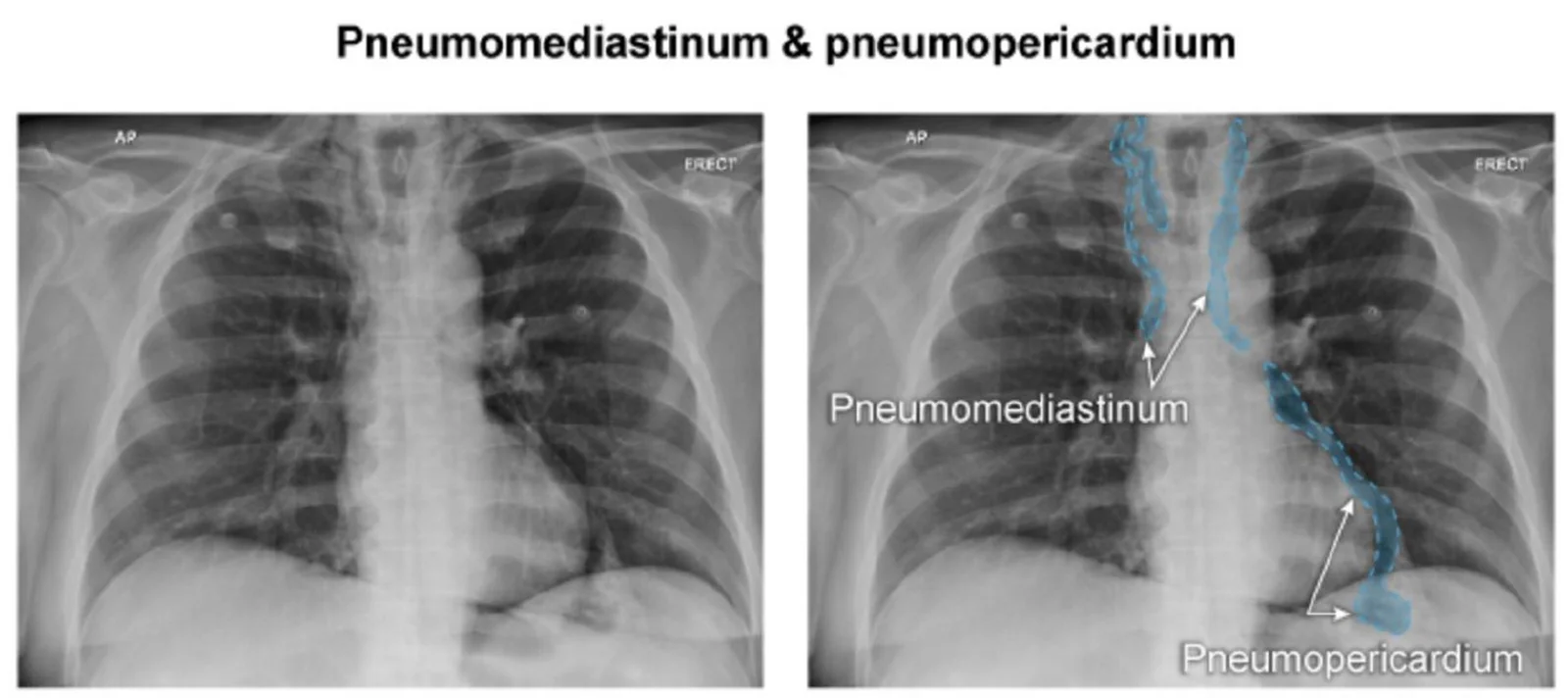
What this shows
A widened mediastinum is a finding, not a diagnosis
tracheobronchial disruption also widens it, but with pneumomediastinum, pneumothorax or subcutaneous emphysema.| Cause | Distinguishing clues | Next step |
|---|---|---|
| Aortic injury | Deceleration, abnormal knob, left hemothorax, pulse asymmetry | CT angiography; TEE if unstable |
| Tracheobronchial rupture | Pneumomediastinum, subcutaneous emphysema, persistent air leak despite a chest tube | Bronchoscopy |
| Esophageal rupture | Pneumomediastinum, effusion, mediastinitis | Contrast esophagram or CT |
| Sternal or venous bleeding | Sternal fracture; normal aorta on CT | Observation |
| Supine film | Magnification, no other findings | Upright film or CT |
Management
Primary survey (airway, breathing, circulation) and stabilization first
then surgical repair.- Other life-threatening injuries (a ruptured spleen, an intracranial bleed) may be treated first while the contained aortic injury is controlled medically.
Anti-impulse therapy while awaiting repair
an intravenous beta blocker (esmolol) to keep the systolic pressure under 100 mm Hg and the heart rate under 80, to prevent extension and rebleeding.The hypotensive patient
resuscitate only to a systolic of about 90 to 100 (permissive hypotension) unless a head injury demands more.Thoracic endovascular aortic repair (TEVAR)
preferred for most injuries (lower mortality and paraplegia than open repair); minimal intimal tears are managed medically with surveillance CT.Complications
free rupture and exsanguination, pseudoaneurysm expansion, paraplegia from spinal cord ischemia, left recurrent laryngeal nerve palsy, endoleak on follow-up.After a high-speed head-on crash, a driver has interscapular pain and a blood pressure of 150/90; the chest film shows a widened mediastinum and a left effusion. What is the next test, and what blood pressure target applies while awaiting repair?
Suspected traumatic aortic injury at the isthmus: he is stable, so CT angiography (TEE if unstable). While awaiting repair (usually TEVAR), an intravenous beta blocker (esmolol) keeps the systolic under 100 and the heart rate under 80.
How it's tested
An unrestrained driver in a head-on collision has a widened mediastinum and an obscured aortic knob; BP 130/80: blunt aortic injury — CT angiography, esmolol to SBP < 100, then TEVAR.
Same patient, BP 70/40 and unstable: TEE in the operating room; emergency repair.
Fall from a roof, widened mediastinum, subcutaneous emphysema and a large persistent air leak from the chest tube: tracheobronchial disruption, not the aorta — bronchoscopy.
Where does the tear occur and why: the isthmus just distal to the left subclavian — the junction of the mobile arch and the fixed descending aorta.
Trauma patient with a contained aortic injury and a subdural hematoma: manage the aorta medically with beta blockade and evacuate the hematoma first; repair the aorta afterwards.
Go deeper
Guidelines: 2022 ACC/AHA Aortic Disease Guideline · ACS Trauma Quality Programs Best Practices Guidelines
Related Step 2 pages: Approach to Aortic Syndromes, Aortic Dissection, Blunt Cardiac Injury, Vascular Trauma, Hypovolemic & Hemorrhagic Shock
High-yield images4
Flashcards for this page
Card 1 of 4 · try-out only, nothing is saved
You just read one page of 236
The rest of Step 2 CK, written exactly like this.
Rapid Review is the reading layer of a full Step 1 platform: a schedule fitted to your exam date, flashcards for every page, and a mentor a message away.
Every Step 1 system
Written the same way: what to know, how it’s tested, where to go deeper.
A plan built to your exam date
Tell us when you sit, and the schedule fits the whole library and your question bank to it.
Flashcards for every page
Ready-made spaced-repetition decks linked to each topic, so nothing you read gets forgotten.
