
Rapid Review·Cardiovascular
Select any text to highlight it or add a note.
Cardiomyopathy
T1Must knowArrhythmogenic Right Ventricular Cardiomyopathy
Focus on
A young adult — often an athlete — with palpitations, syncope or cardiac arrest from a VT with a left bundle branch block morphology, an epsilon wave and T inversions in V1–V3, and a dilated, fibrofatty right ventricle on MRI. Beta blocker, ICD, no competitive sport.
Key takeaways
What it is
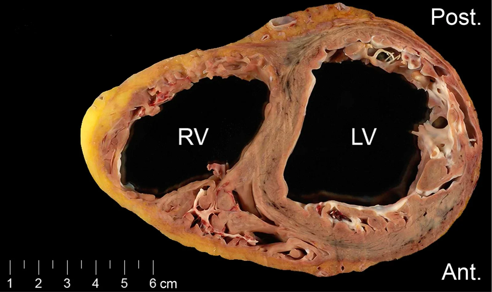
What this shows
A genetic cardiomyopathy of the right ventricle
non-ischemic, and associated with ventricular arrhythmias.Fibrofatty replacement
myocyte death with fibrofatty replacement thins and dilates the right ventricular wall.- The damaged wall then generates arrhythmias and fails.
Clinical features
The young adult who collapses
cardiogenic syncope or sudden cardiac death in a young adult.Diagnosis
Imaging and genes
echocardiography, cardiac magnetic resonance imaging (MRI), and genetic testing.Electrocardiogram (ECG)
three findings.- Repolarization abnormalities in the right precordial leads (V1 to V3).
- Ventricular arrhythmias.
- Sometimes an epsilon wave (a small notch just after a widened QRS, before the T wave), which is specific but not sensitive.
Management
Antiarrhythmic drugs
with a beta blocker first line.Implantable cardioverter-defibrillator (ICD)
for those at risk of sudden death.Avoid competitive sports.
A 22-year-old runner faints during a race. The ECG shows T-wave inversions in V1 to V3 and a small notch just after the QRS. What is the notch called, what does MRI show, and what is first-line drug therapy?
An epsilon wave, in arrhythmogenic right ventricular cardiomyopathy. MRI shows a thin, dilated right ventricle with fibrofatty replacement. First line is a beta blocker, with an ICD for those at risk of sudden death and no competitive sports.
How it's tested
A 26-year-old marathoner faints during a race; the ECG afterwards shows T-wave inversions in V1–V3 and a small notch after the QRS in V1; MRI shows a dilated RV with fatty infiltration: ARVC — ICD, beta blocker, and no more competitive endurance sport.
Wide-complex tachycardia with LBBB morphology in a young adult with a family history of sudden death: think RV origin — ARVC until the heart is proven structurally normal.
Same VT morphology in a young woman with a completely normal heart on MRI, provoked by exercise: idiopathic RVOT tachycardia — benign; beta blocker or ablation.
Go deeper
Guidelines: 2023 ESC Cardiomyopathy Guideline
Related Step 2 pages: Dilated Cardiomyopathy, Ventricular Tachycardia, Brugada Syndrome, Cardiac Implantable Devices, Approach to Syncope
High-yield images1
You just read one page of 236
The rest of Step 2 CK, written exactly like this.
Rapid Review is the reading layer of a full Step 1 platform: a schedule fitted to your exam date, flashcards for every page, and a mentor a message away.
Every Step 1 system
Written the same way: what to know, how it’s tested, where to go deeper.
A plan built to your exam date
Tell us when you sit, and the schedule fits the whole library and your question bank to it.
Flashcards for every page
Ready-made spaced-repetition decks linked to each topic, so nothing you read gets forgotten.
