
Rapid Review·Cardiovascular
Select any text to highlight it or add a note.
Cardiac Trauma
T1Must knowBlunt Cardiac Injury
Focus on
After a steering-wheel impact or a fall, an ECG and a troponin decide who needs cardiac monitoring: both normal rules out significant injury; either abnormal means telemetry. A sternal fracture on its own does not. Echo is for the unstable patient or the persistent arrhythmia — to find contusion, tamponade, a septal or valve rupture, or a torn coronary.
Key takeaways
What it is and what causes it
Blunt cardiac injury
myocardial injury from blunt chest trauma or sudden deceleration (the heart compressed between sternum and spine, or a direct precordial blow).- Injured myocytes and pacemaker cells give reduced contractility and arrhythmias.
A spectrum
from silent troponin release and transient arrhythmia to fatal rupture.- Myocardial contusion: patchy hemorrhage and necrosis with contractile dysfunction.
- Coronary dissection (traumatic infarction), valvular or septal rupture, free-wall rupture with tamponade.
- Commotio cordis: ventricular fibrillation from a precordial blow during repolarization.
Right-sided injuries are the most common
the right atrium and ventricle sit against the chest wall (Cardiac Anatomy).Causes
motor vehicle collisions (steering wheel, unrestrained), falls, crush, sports (a baseball to the chest in a child), resuscitation compressions.- Companions: sternal and rib fractures, pulmonary contusion, hemothorax and aortic injury (Traumatic Aortic Injury).
Clinical features
Range
from asymptomatic arrhythmias to rapidly fatal rupture; chest-wall pain is universal and unhelpful.Suggestive findings
persistent unexplained tachycardia despite fluids once hemorrhage is excluded, new arrhythmia (atrial fibrillation, ectopy, ventricular tachycardia, a new bundle branch block or conduction delay), hypotension not explained by hemorrhage.- A new S3 or murmur (valve or septal rupture), tamponade signs, ischemic ST elevation (coronary injury).
A sternal fracture alone
does not diagnose or predict cardiac injury; mechanism and physiology guide screening.The screening pathway
Electrocardiogram plus troponin in every patient with a suggestive mechanism or findings
per the Blunt chest trauma pathway.Both normal
clinically significant injury is excluded (no cardiac monitoring for this alone).Any new electrocardiographic abnormality or a raised troponin
admit for continuous monitoring 24 to 48 hours with serial troponin.- New abnormalities include atrial fibrillation, T-wave inversion, block, unexplained sinus tachycardia.
- Troponin rises are usually mild and do not grade severity.
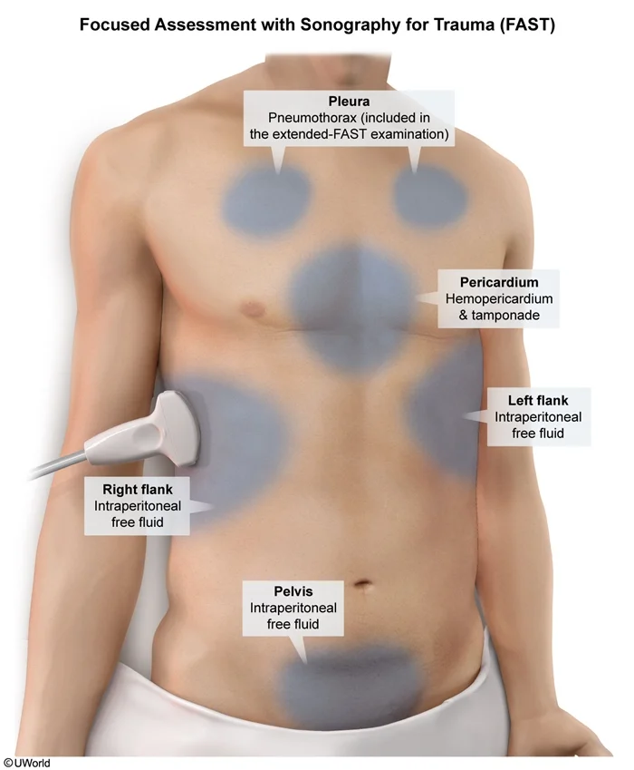
What this shows
Echocardiography is not a screening test
obtain it immediately for hemodynamic instability, persistent arrhythmia, a new murmur or heart failure.- It shows regional or global hypokinesis (often the right ventricle), reduced ejection fraction, effusion or tamponade, septal defect, valve disruption, wall thrombus.
ST elevation or a large territorial wall-motion abnormality
coronary computed tomography (CT) angiography or catheterization to separate contusion from traumatic coronary occlusion.- Thrombolysis is contraindicated in the multiply injured.
| Cause | Neck veins and lungs | Bedside clue | Response |
|---|---|---|---|
| Ongoing hemorrhage | Flat neck veins, clear lungs | Positive trauma ultrasound, pelvis, long bones, chest-tube output; falling hemoglobin | Blood products and source control (Hypovolemic & Hemorrhagic Shock) |
| Cardiac Tamponade | Distended neck veins, clear lungs | Effusion with chamber collapse, pulsus paradoxus | Pericardiocentesis or thoracotomy |
| Tension pneumothorax | Distended neck veins, unilateral absent breath sounds | Clinical; absent lung sliding | Needle decompression, chest tube |
| Myocardial contusion | Neck veins raised, bibasilar crackles | Negative trauma ultrasound, an arrhythmia, a raised wedge pressure that worsens with fluids; echo shows a hypokinetic wall | Inotropes, cautious fluids, monitoring (Cardiogenic Shock) |
| Traumatic coronary occlusion | Variable | Regional ST elevation, territorial wall-motion abnormality | Coronary imaging, percutaneous intervention |
| Neurogenic shock | Flat neck veins, warm skin, bradycardia | Spinal cord injury | Fluids then vasopressors (Distributive Shock) |
Management and complications
Serial electrocardiogram monitoring after admission
for progression of changes or arrhythmias.- Most arrhythmias are transient, treated by standard protocols after correcting hypoxia, hypovolemia, electrolytes, acidosis and pain; no prophylactic antiarrhythmics.
Contusion with cardiogenic dysfunction
cautious monitored fluid, a small bolus with reassessment (the injured ventricle is stiff and overloads easily).- Inotropes (dobutamine, milrinone) for low output, vasopressors for pressure, guided by echo or invasive hemodynamics.
Structural injuries get specific treatment
drainage or thoracotomy for tamponade, emergency surgery for septal, valvular or free-wall rupture, percutaneous intervention (no lytics) for a torn coronary when other bleeding allows, pacing for high-grade block.Commotio cordis
immediate defibrillation and resuscitation.Anticoagulation
avoid until intracranial and solid-organ bleeding are excluded.Recovery
most contusions recover within days to weeks; follow-up echocardiography if dysfunction was found.Complications
malignant arrhythmias (including delayed ventricular fibrillation) and cardiogenic shock (profound hypotension with normal volume, bibasilar crackles, raised jugular pressure and wedge pressure).- Right-ventricular failure, tamponade, delayed rupture or pseudoaneurysm, valve regurgitation (tricuspid most often), septal defect, mural thrombus with embolism, and post-cardiac injury syndrome weeks later (Post-Cardiac Injury Syndrome).
After a steering-wheel injury, a hypotensive driver has a negative FAST, bibasilar crackles, a new arrhythmia and a wedge pressure that rises further after fluids. What is the diagnosis and the treatment?
Myocardial contusion with cardiogenic shock (blunt cardiac injury). Stop the fluids, support with inotropes (dobutamine, milrinone) and vasopressors as needed, monitor continuously, and get an echocardiogram (hypokinetic wall, often the right ventricle).
How it's tested
A restrained driver with steering-wheel bruising and a sternal fracture; ECG and troponin are normal: significant blunt cardiac injury is excluded — no telemetry is needed for the heart; the fracture alone does not mandate it.
Same patient with new frequent PVCs and a troponin of 0.4: admit for continuous monitoring and serial troponin; echo if arrhythmia persists or he becomes hypotensive.
Persistent hypotension after hemorrhage has been controlled, rising CVP, and worsening with each fluid bolus: echocardiography — myocardial contusion with ventricular dysfunction vs tamponade (Blunt Cardiac Injury).
ST elevation in V1–V4 after a motorcycle crash: traumatic LAD dissection or thrombosis until proven otherwise — coronary imaging; PCI rather than thrombolysis.
A 12-year-old collapses after a baseball strikes his chest: commotio cordis — ventricular fibrillation; CPR and immediate defibrillation.
A motor-vehicle-crash victim remains hypotensive after two units of blood with a negative FAST and a stable pelvis; he has frequent PVCs and a new RBBB, and his pressure falls further after a liter of saline; echo shows a hypokinetic right ventricle without effusion: myocardial contusion with cardiogenic dysfunction — inotropic support and monitoring, not more fluid.
Which chamber is most often contused and why: the right ventricle — it lies directly behind the sternum.
Troponin of 2.0 after chest trauma with a normal echo and no arrhythmia: monitor 24–48 hours; the troponin value alone does not grade severity or mandate intervention.
Contusion patient develops ST elevation in V2–V4 with a new anterior wall-motion abnormality: traumatic LAD injury — coronary angiography and PCI, no thrombolytics.
Go deeper
Guidelines: EAST Practice Management Guideline — Screening for Blunt Cardiac Injury
Related Step 2 pages: Blunt Cardiac Injury, Cardiac Tamponade, Traumatic Aortic Injury, Acute Coronary Syndrome, Vascular Trauma, Cardiac Anatomy
High-yield images2
Flashcards for this page
Card 1 of 3 · try-out only, nothing is saved
You just read one page of 236
The rest of Step 2 CK, written exactly like this.
Rapid Review is the reading layer of a full Step 1 platform: a schedule fitted to your exam date, flashcards for every page, and a mentor a message away.
Every Step 1 system
Written the same way: what to know, how it’s tested, where to go deeper.
A plan built to your exam date
Tell us when you sit, and the schedule fits the whole library and your question bank to it.
Flashcards for every page
Ready-made spaced-repetition decks linked to each topic, so nothing you read gets forgotten.
