
Rapid Review·Cardiovascular
Select any text to highlight it or add a note.
Pericardial Disease
T1Must knowCardiac Tamponade
Focus on
Fluid under pressure stops the heart from filling: hypotension, distended neck veins, muffled sounds (Beck triad), tachycardia and pulsus paradoxus, with chamber collapse on echo. Fluids buy minutes; the needle (or the surgeon, in trauma and dissection) is the treatment. Positive-pressure ventilation and diuretics make it worse.
Key takeaways
What it is
Impaired diastolic filling
because a pericardial effusion is under pressure and compresses the heart, especially the right ventricle.Two ways to get there
a large effusion (over 2 L) or the rapid accumulation of a smaller one.| Acute | Subacute | |
|---|---|---|
| Speed of accumulation | Minutes to hours | Days to weeks |
| Effusion volume | 100 to 200 mL | 1 to 2 L |
| Chest radiograph | Normal silhouette | Enlarged, globular silhouette |
Clinical features
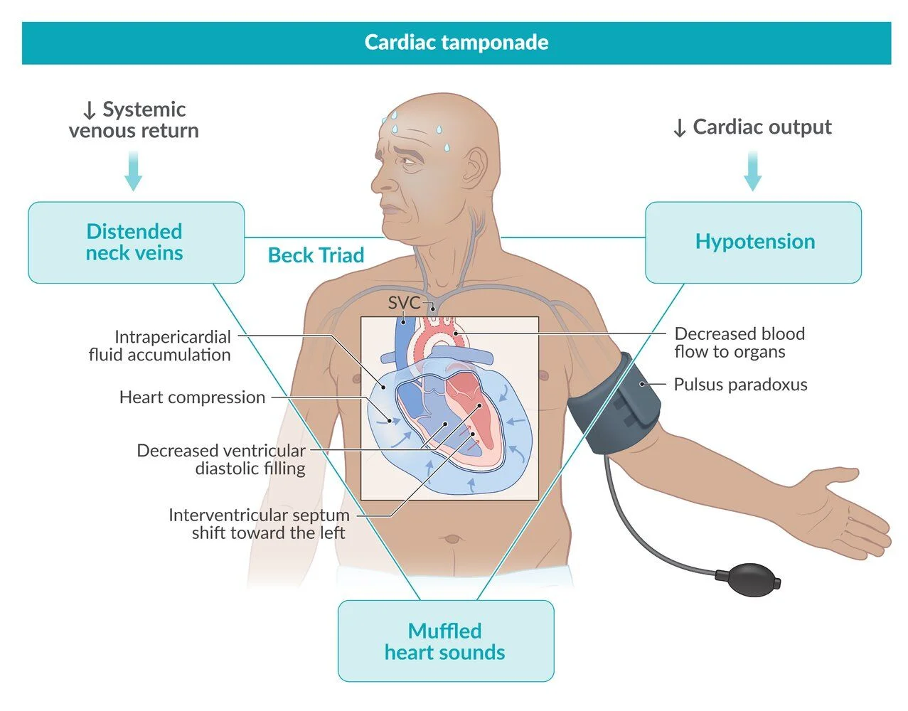
What this shows
Presentation
chest pain, dyspnea and hemodynamic compromise.Beck triad
hypotension, jugular venous distension, muffled heart sounds.Other signs
pulsus paradoxus (systolic blood pressure falls by more than 10 mm Hg on inspiration), a narrow pulse pressure, distant heart sounds.It mimics cardiogenic shock without pulmonary edema.
Slow effusions look different
a slowly accumulating large effusion gives the globular silhouette with clear lungs seen in the Large pericardial effusion on chest radiograph.Diagnosis
Unstable patient
investigate only when the patient is stable enough; an unstable patient with a typical picture goes straight to treatment.Transthoracic echocardiography
confirms the effusion.- Right atrial and ventricular collapse in early diastole.
- A plethoric inferior vena cava that does not collapse on inspiration.
- Equalization of diastolic pressures across the four chambers with a low cardiac output.
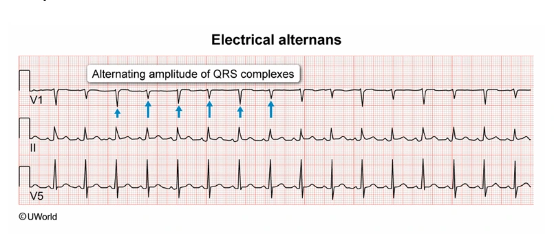
What this shows
Electrocardiogram (ECG)
low-voltage QRS (non-specific) and electrical alternans, which is more specific.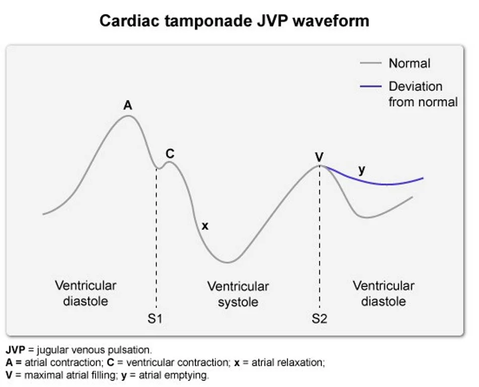
What this shows
Jugular venous tracing
a blunted or absent y descent, because the ventricles barely fill.Management
Intravenous fluids cautiously
to support preload.Non-hemorrhagic tamponade
emergency pericardiocentesis.Hemorrhagic (traumatic) tamponade
emergency pericardiocentesis followed by a pericardial window.- A window is also used for loculated, malignant or rapidly recurrent effusions.
Do not delay surgery
use a pericardial window when pericardiocentesis is unlikely to hold because the fluid reaccumulates at once.Pulsus paradoxus and Kussmaul sign
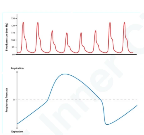
What this shows
Pulsus paradoxus
a fall in pulse amplitude and systolic blood pressure of more than 10 mm Hg on inspiration.- Seen with constrictive cardiac disease (tamponade, tension pneumothorax) and with severe obstructive lung disease (chronic obstructive pulmonary disease (COPD), asthma).
- For the exam, constrictive pericarditis does not cause it.
Kussmaul sign
a paradoxical rise in jugular venous pressure on inspiration.- Normally inspiration lowers intrathoracic pressure, pulls venous blood into the right ventricle, and lowers the jugular pressure.
- When the right ventricle cannot accept that extra return, blood backs up into the venae cavae and the neck veins rise.
- Seen in constrictive pericarditis, restrictive cardiomyopathy, right heart failure, massive pulmonary embolism, and right-sided tumors.
| Sign | Cardiac tamponade | Restrictive cardiomyopathy | Constrictive pericarditis |
|---|---|---|---|
| Pulsus paradoxus | Yes | No | No |
| Kussmaul sign | No | Yes | Yes |
After a stab wound to the chest, a patient is hypotensive with distended neck veins, muffled heart sounds and clear lungs; the systolic pressure drops 18 mm Hg on inspiration. What is the diagnosis, what should be avoided, and what is the treatment?
Cardiac tamponade (Beck triad plus pulsus paradoxus). Give IV fluids cautiously and avoid positive-pressure ventilation. Treat a traumatic (hemorrhagic) tamponade with emergency pericardiocentesis followed by a pericardial window.
How it's tested
Stab wound to the left chest with BP 70/40, distended neck veins and muffled sounds: tamponade — emergency thoracotomy/surgical drainage; pericardiocentesis only as a bridge if arrest is imminent.
Tearing chest pain, hypotension, JVD and pulsus paradoxus: dissection ruptured into the pericardium — emergency aortic surgery; no routine pericardiocentesis.
Cancer patient with dyspnea, tachycardia, BP 85/60, electrical alternans and a large effusion with RV collapse: echo-guided pericardiocentesis now; a window later because it will recur.
Post-CABG day 1: shock with equalized diastolic pressures and no obvious circumferential effusion: localized clot compressing the heart — surgical re-exploration.
Severe asthma attack with a 15 mm Hg pulsus paradoxus and no effusion on echo: pulsus paradoxus is not specific — treat the asthma.
Go deeper
Guidelines: 2025 ESC Myocarditis and Pericarditis Guideline
Related Step 2 pages: Acute Pericarditis, Pericardial Effusion, Cardiac Tamponade, Obstructive Shock, Aortic Dissection, Post MI Complications, Blunt Cardiac Injury
High-yield images5
Flashcards for this page
Card 1 of 5 · try-out only, nothing is saved
You just read one page of 236
The rest of Step 2 CK, written exactly like this.
Rapid Review is the reading layer of a full Step 1 platform: a schedule fitted to your exam date, flashcards for every page, and a mentor a message away.
Every Step 1 system
Written the same way: what to know, how it’s tested, where to go deeper.
A plan built to your exam date
Tell us when you sit, and the schedule fits the whole library and your question bank to it.
Flashcards for every page
Ready-made spaced-repetition decks linked to each topic, so nothing you read gets forgotten.
