
Rapid Review·Cardiovascular
Select any text to highlight it or add a note.
Pericardial Disease
T2High yieldConstrictive Pericarditis
Focus on
A rigid scarred pericardium that stops the heart from filling: chronic right-sided failure with a JVP that rises on inspiration, a sharp y descent, a pericardial knock, and calcification on the film — after tuberculosis, radiation, or cardiac surgery. The exam pairs it with restrictive cardiomyopathy every time; pericardiectomy cures constriction.
Key takeaways
What it is and what causes it
A rigid shell around the heart
fibrous scarring makes the pericardium rigid and thick, so it restricts diastolic filling.United States
idiopathic or recurrent viral pericarditis is the most common cause; prior cardiac surgery or radiation is second.Developing world
tuberculosis is very common.Other
connective tissue and autoimmune disease.Clinical features
Prominent right heart failure
fatigue, exertional dyspnea, tachycardia, a raised jugular venous pressure, peripheral edema, ascites.Kussmaul sign
the jugular pressure rises with inspiration.The liver and gut suffer too
from the high venous pressure.- Congestive hepatopathy with hepatomegaly that can progress to cardiac cirrhosis.
- Hypoalbuminemia from a protein-losing enteropathy (intestinal lymphangiectasia from the high venous pressure).
Pericardial knock
a high-pitched early diastolic sound like a premature S3.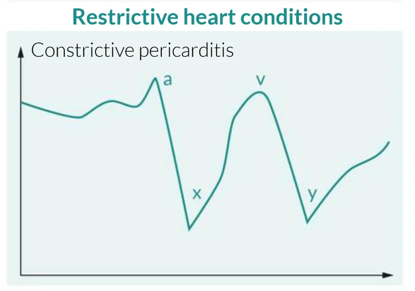
What this shows
Prominent x and y descents
on the venous pressure tracing.Diagnosis
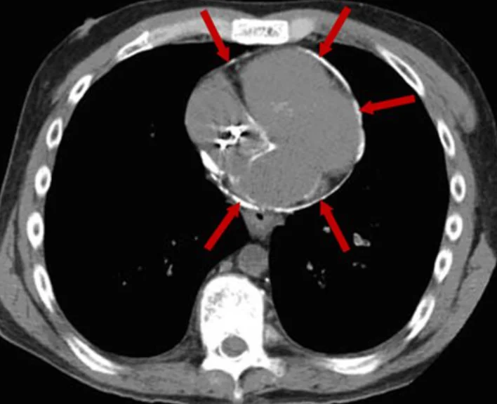
What this shows
Chest radiograph
pericardial thickening and sometimes calcification.Echocardiography is confirmatory
three findings.- Bi-atrial enlargement.
- Normal ventricular wall thickness and cavity size.
- A septum that shifts leftward on inspiration.
Electrocardiogram
low-voltage QRS.Cardiac magnetic resonance imaging (MRI) or computed tomography (CT)
shows the thickened pericardium and the extent of calcification.Management
Supportive care; pericardiectomy is definitive
surgical removal of the pericardium relieves the constriction.Avoid beta blockers and calcium-channel blockers
they slow the compensatory tachycardia and worsen the failure.Sedatives or positive-pressure ventilation
can cause hemodynamic collapse.A man treated with chest radiation years ago has ascites, edema, a raised jugular venous pressure that rises on inspiration and an early diastolic knock; CT shows a calcified pericardium. What is the definitive treatment, and which rate-slowing drugs are harmful?
Constrictive pericarditis (Kussmaul sign, pericardial knock, calcification). Pericardiectomy is definitive. Beta blockers and calcium-channel blockers are harmful because they slow the compensatory tachycardia that keeps the output up.
How it's tested
A man treated with mediastinal radiation for Hodgkin lymphoma 15 years ago has edema, ascites, a JVP that rises on inspiration and an early diastolic knock: constrictive pericarditis — CT/echo, then pericardiectomy.
JVP with a sharp y descent that rises on inspiration: Kussmaul sign plus prominent y descent = constriction; tamponade blunts the y descent and lacks Kussmaul sign.
Echo shows a septal bounce, preserved medial e′ and 40% respiratory variation in mitral inflow; walls are normal thickness: constriction, not restrictive cardiomyopathy.
Simultaneous catheterization: with inspiration the RV systolic pressure rises as the LV systolic pressure falls: ventricular discordance — constriction.
Persistent edema and ascites despite escalating diuretics: pericardiectomy — diuretics cannot remove a mechanical shell.
Go deeper
Guidelines: 2025 ESC Myocarditis and Pericarditis Guideline
Related Step 2 pages: Acute Pericarditis, Restrictive Cardiomyopathy, Cardiac Tamponade, Jugular Venous Pressure
High-yield images2
Flashcards for this page
Card 1 of 5 · try-out only, nothing is saved
You just read one page of 236
The rest of Step 2 CK, written exactly like this.
Rapid Review is the reading layer of a full Step 1 platform: a schedule fitted to your exam date, flashcards for every page, and a mentor a message away.
Every Step 1 system
Written the same way: what to know, how it’s tested, where to go deeper.
A plan built to your exam date
Tell us when you sit, and the schedule fits the whole library and your question bank to it.
Flashcards for every page
Ready-made spaced-repetition decks linked to each topic, so nothing you read gets forgotten.
