
Rapid Review·Cardiovascular
Select any text to highlight it or add a note.
Cardiomyopathy
T1Must knowDilated Cardiomyopathy
Focus on
A big, weak heart: EF < 40% with dilated chambers, an S3 and functional MR. The cause list is the exam — alcohol (reversible with abstinence), doxorubicin (irreversible) vs trastuzumab (reversible), viral myocarditis, Chagas, peripartum, tachycardia, wet beriberi, hemochromatosis — and treatment is HFrEF therapy plus removing the cause.
Key takeaways
Cardiomyopathies at a glance
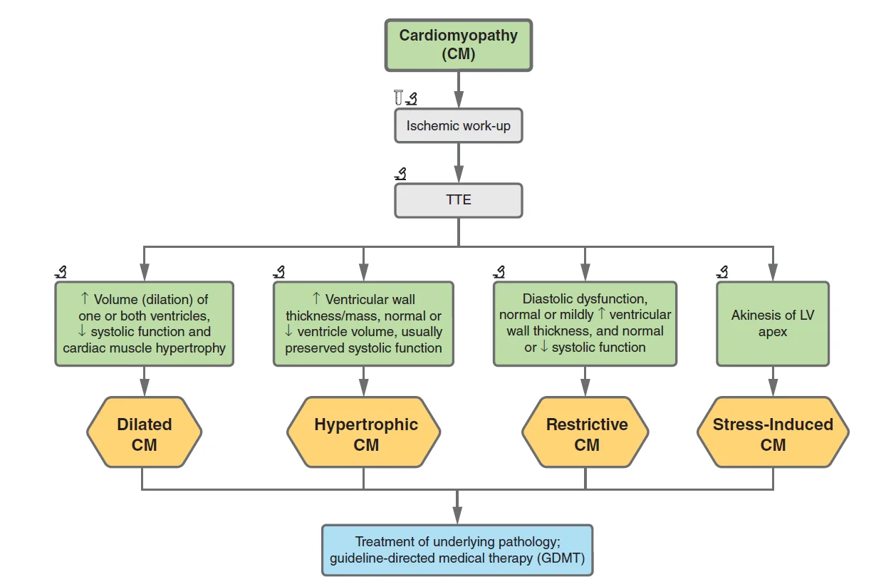
What this shows
A cardiomyopathy
a disease of the heart muscle, acquired or inherited, that impairs pumping and leads to arrhythmias and heart failure.Dilated cardiomyopathy (DCM)
dilation with impaired contraction of one or both ventricles, with an ejection fraction (EF) under 40 percent.Eccentric hypertrophy
sarcomeres are added in series, so the chambers dilate.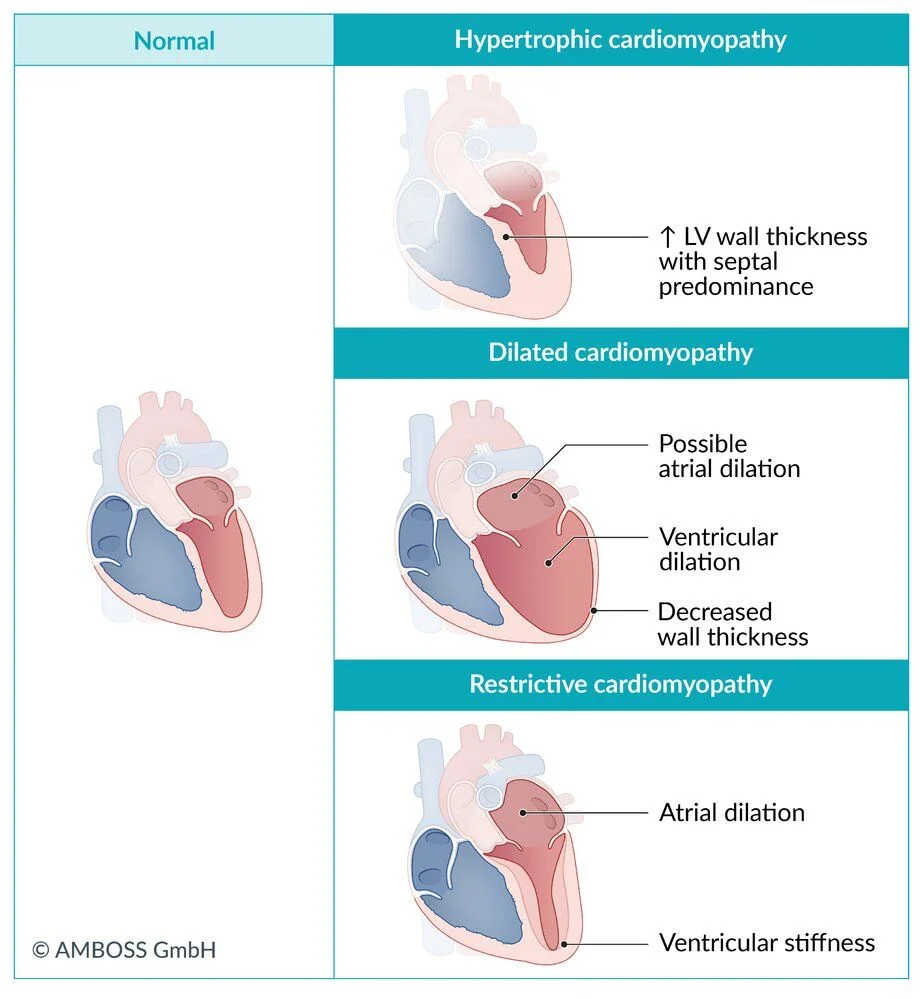
What this shows
| Type | Mechanism | Causes | Findings |
|---|---|---|---|
| Dilated (DCM) | Systolic dysfunction; eccentric hypertrophy (sarcomeres in series) | Idiopathic or familial (TTN gene); ischemic heart disease (most common secondary), hypertension, alcohol, myocarditis, drugs (doxorubicin, zidovudine, cocaine), peripartum, radiation, infections (coxsackievirus, HIV, Chagas), endocrine disease, wet beriberi | S3, systolic regurgitant murmur; "balloon" heart on chest radiograph; dilated chambers on echo |
| Hypertrophic (HCM) | Diastolic dysfunction; concentric hypertrophy (sarcomeres in parallel) | Hypertension (most common acquired), aortic stenosis, Friedreich ataxia, hypertrophic obstructive cardiomyopathy (HOCM) | S4, systolic murmur with mitral regurgitation; left ventricular hypertrophy with septal Q waves; septal hypertrophy, systolic anterior motion of the mitral valve, outflow obstruction on echo |
| Restrictive (RCM) | Diastolic dysfunction without systolic dysfunction; normal or near-normal EF | Infiltration (amyloidosis, hemochromatosis, sarcoidosis), scleroderma, Loeffler eosinophilic endocarditis, endomyocardial fibrosis, radiation | Right-sided failure predominates (jugular distension, edema, ascites); low voltage or left bundle branch block; rapid early filling on echo; biopsy may show the infiltrate |
| Stress-induced (Takotsubo) | Transient apical ballooning from catecholamine surge | Marked physical or emotional stress | Looks like an infarct (chest pain, ST elevation, troponin) with normal coronaries; mid and apical hypokinesis with basal hyperkinesis |
Causes of dilated cardiomyopathy
Primary
idiopathic or familial, most often a mutation in the TTN gene (titin).Ischemic heart disease
the most common secondary cause.- It gives focal wall-motion abnormalities, whereas a non-ischemic DCM gives global hypokinesis.
Infection
viral myocarditis (coxsackievirus, HIV), Chagas disease, Lyme disease.Systemic disease
long-standing poorly controlled hypertension, endocrine disease (thyroid disease, acromegaly, pheochromocytoma), late hemochromatosis, sarcoidosis.Nutritional
thiamine deficiency (wet beriberi).Peripartum
the last month of pregnancy or the first five months after delivery (Peripartum Cardiomyopathy).Toxic
alcohol, chemotherapy, zidovudine, cocaine, radiation.Tachycardia-mediated
a chronically rapid rate, such as untreated atrial fibrillation with a rapid ventricular response.Clinical features and diagnosis
Systolic heart failure.
Auscultation
an S3, and a systolic regurgitant murmur from the dilated mitral or tricuspid rings.Transthoracic echocardiography (TTE)
the gold standard imaging test; shows dilated chambers.- The full workup by cause is on the DCM workup.
Chest radiograph
a "balloon" appearance of the heart.Management
Treat as heart failure with reduced ejection fraction
(Heart Failure) guideline-directed therapy.- A beta blocker, an angiotensin-system inhibitor, a mineralocorticoid receptor antagonist and a sodium-glucose cotransporter-2 (SGLT2) inhibitor.
Diuretics
first line for symptoms of volume overload.Treat the cause
two classic examples.- Abstinence is the mainstay for alcoholic cardiomyopathy.
- Rate or rhythm control is the initial treatment for tachycardia-mediated cardiomyopathy.
A woman treated for breast cancer develops a dilated cardiomyopathy. Which chemotherapy drug makes recovery likely when it is stopped, and which one does not?
Trastuzumab damage is not structural and usually reverses fully when the drug is stopped. Doxorubicin damage is irreversible.
How it's tested
A 45-year-old man drinking a bottle of whiskey daily presents with heart failure and an EF of 20% with global hypokinesis: alcoholic dilated cardiomyopathy — abstinence plus HFrEF drugs; function often recovers.
A breast-cancer patient develops heart failure after her anthracycline course: doxorubicin cardiomyopathy — dose-dependent and usually irreversible; trastuzumab-related dysfunction would be expected to recover.
Persistent AF with ventricular rates of 130–150 for months, now EF 30%: tachycardia-mediated cardiomyopathy — rate control or ablation first; the EF improves.
Immigrant from Brazil with heart failure, an apical LV aneurysm and dysphagia: Chagas cardiomyopathy.
Alcoholic with heart failure, warm extremities and high output: wet beriberi — thiamine before glucose.
Go deeper
Guidelines: 2023 ESC Cardiomyopathy Guideline · 2022 AHA/ACC/HFSA Heart Failure Guideline
Related Step 2 pages: Dilated Cardiomyopathy, Heart Failure, Myocarditis, Peripartum Cardiomyopathy, Atrial Fibrillation, Cardiac Implantable Devices
High-yield images3
Flashcards for this page
Card 1 of 5 · try-out only, nothing is saved
You just read one page of 236
The rest of Step 2 CK, written exactly like this.
Rapid Review is the reading layer of a full Step 1 platform: a schedule fitted to your exam date, flashcards for every page, and a mentor a message away.
Every Step 1 system
Written the same way: what to know, how it’s tested, where to go deeper.
A plan built to your exam date
Tell us when you sit, and the schedule fits the whole library and your question bank to it.
Flashcards for every page
Ready-made spaced-repetition decks linked to each topic, so nothing you read gets forgotten.
