
Rapid Review·Cardiovascular
Select any text to highlight it or add a note.
Heart Failure
T1Must knowHeart Failure
Focus on
Recognize the syndrome (orthopnea, PND and an S3 are the specific findings; dyspnea and BNP the sensitive ones), confirm with echo and classify by ejection fraction, then treat the acute decompensation by phenotype — loop diuretic if congested, nitrates and positive pressure for hypertensive flash edema, inotropes only for shock — before building chronic therapy.
Key takeaways
What heart failure is
A clinical syndrome
heart failure (HF) comes from a cardiomyopathy that impairs ventricular filling or ejection, so the heart cannot pump blood effectively.- Congestive heart failure is HF with signs of fluid overload, such as peripheral edema or jugular venous distension.
Left vs right
left heart failure causes tissue hypoperfusion and raised pulmonary capillary pressure; right heart failure raises central venous pressure.- One-sided failure often becomes biventricular.
Systolic vs diastolic dysfunction
two different mechanical problems.- Systolic: reduced contractility with ventricular enlargement and a low ejection fraction.
- Diastolic: a stiff, poorly relaxing ventricle with high filling pressures.
| Subtype | Ejection fraction | Mechanism | Causes |
|---|---|---|---|
| HF with reduced ejection fraction (HFrEF) | Under 40 percent | Systolic dysfunction | Dilated cardiomyopathy (ischemic or non-ischemic), myocarditis |
| HF with mildly reduced ejection fraction (HFmrEF) | 40 to 49 percent | Intermediate | As above |
| HF with preserved ejection fraction (HFpEF) | Over 50 percent | Diastolic dysfunction | Hypertension (the number one cause), obesity and a sedentary lifestyle, diabetes, aging, hypertrophic and restrictive cardiomyopathy |
Causes
the three major causes are coronary artery disease (CAD, the most common), hypertension and diabetes mellitus.- Patients usually have several risk factors together.
Clinical features
Sensitive vs specific findings
dyspnea and a raised B-type natriuretic peptide (BNP) are the most sensitive; orthopnea, paroxysmal nocturnal dyspnea and an S3 gallop are the most specific.- In a patient over 40, an S3 gallop is the most specific finding for decompensated HF.
The gallop tells the subtype
an S3 points to HFrEF (volume overload); an S4 points to HFpEF (ventricular hypertrophy).Common to both sides
fatigue, nocturia (edema fluid mobilizes when the patient lies flat at night), tachycardia and arrhythmias.- Pulsus alternans: alternating strong and weak pulses with a regular rhythm, from a beat-to-beat change in cardiac output.
Left-sided
pulmonary congestion.- Pulmonary edema, dyspnea, orthopnea, paroxysmal nocturnal dyspnea and cough (worse supine), and cardiac asthma from airway compression.
- Examination: bilateral basal crackles, a laterally displaced point of maximal impulse, and cool pale legs.
- Slowly progressive congestion (over months) may show little or no pulmonary edema, because the pulmonary lymphatics can raise their drainage up to ten-fold.
Right-sided
fluid retention from a high central venous pressure.- Jugular venous distension (jugular venous pressure over 8 cm), peripheral edema, hepatosplenomegaly.
- Kussmaul sign: the neck veins distend on inspiration.
- Hepatojugular reflux: pressing on the liver raises the jugular venous pressure.
Diagnosis and classification
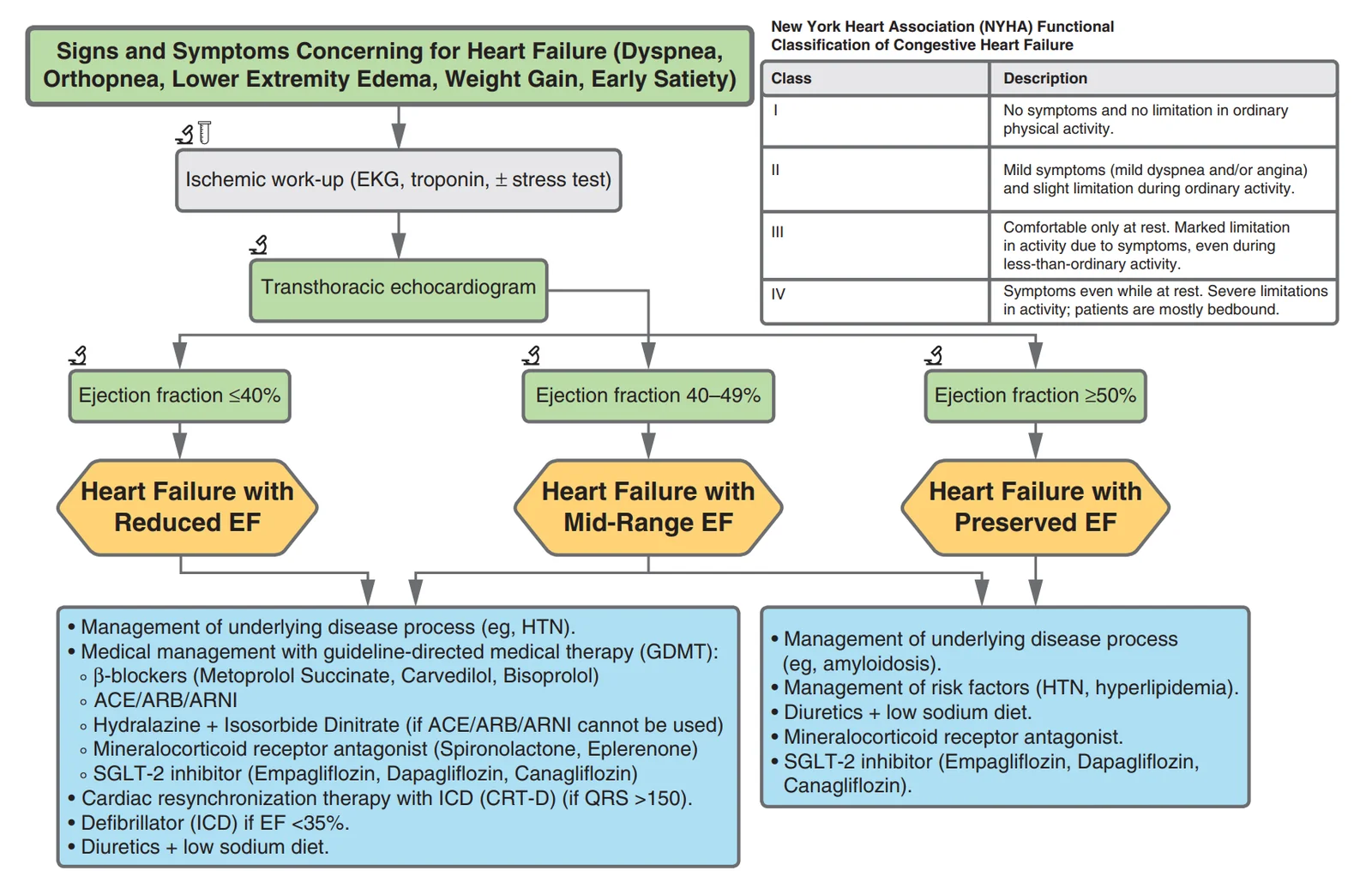
What this shows
The diagnosis is mainly clinical
supported by tests.BNP
the screening test for every patient.- It is highly sensitive, so a low BNP excludes HF.
- It is falsely low in obesity because fat clears it.
Transthoracic echocardiography (TTE)
the initial imaging test; it confirms the diagnosis and measures systolic ejection fraction and diastolic function.Chest radiograph
each finding is a stage of fluid.- Alveolar edema in a "bat's wing" pattern.
- Interstitial edema with Kerley B lines (short septal lines perpendicular to the pleura).
- Cephalization of the pulmonary vessels.
- Pleural effusion, and cardiomegaly (cardiothoracic ratio over 0.5).
Right heart catheterization
a raised pulmonary capillary wedge pressure in left-sided failure; raised right atrial and right ventricular end-diastolic pressures in right-sided failure.| Class | Symptoms |
|---|---|
| I | No limitation; no symptoms of HF with ordinary activity |
| II | Mild symptoms and slight limitation with ordinary activity; comfortable at rest |
| III | Marked limitation; less-than-ordinary activity causes symptoms; comfortable only at rest |
| IV | Symptoms at rest and with any activity |
Lifestyle for every patient
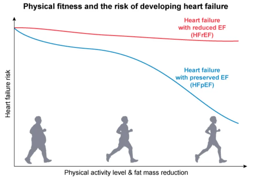
What this shows
Aerobic exercise
brisk walking for at least 150 minutes a week.- Weight loss is especially preventive for HFpEF.
A healthy diet
Dietary Approaches to Stop Hypertension (DASH) or Mediterranean, with dietary sodium under 2 g a day to limit congestion.- Fluid restriction does not reduce hospitalization or mortality.
Smoking cessation
and avoidance of alcohol and recreational drugs.Adherence pays
these measures lower hospitalization and cardiovascular and all-cause mortality.Treating HFrEF
The 4 drugs that improve survival
think "RAAS + BB + MRA + SGLT2" (renin-angiotensin-aldosterone system (RAAS) inhibitor, beta blocker (BB), mineralocorticoid receptor antagonist, sodium-glucose cotransporter-2 (SGLT2) inhibitor).- RAAS inhibitor: preferred is an ARNI, sacubitril/valsartan; if an ARNI cannot be used, an ACEi/ARB.
- Evidence-based β-blocker: only remember these 3: carvedilol, metoprolol (succinate), bisoprolol.
- Mineralocorticoid receptor antagonist (MRA), spironolactone / eplerenone: use if ejection fraction (EF) ≤35% + NYHA II to IV, or after an ST-elevation myocardial infarction (STEMI) with EF ≤40% + HF symptoms or diabetes; avoid if K⁺ >5 mEq/L or CrCl <30 mL/min.
- SGLT2 inhibitor, dapagliflozin / empagliflozin: reduces HF hospitalization and mortality, and works with or without diabetes.
Congested? Add a diuretic
furosemide; if resistant, add metolazone.- Diuretics improve symptoms, not survival.
Still symptomatic
choose by the situation.- Cannot tolerate a RAAS inhibitor: hydralazine + isosorbide dinitrate.
- HFrEF with atrial fibrillation (AF) or persistent symptoms: digoxin.
- Sinus rhythm and heart rate (HR) ≥70 despite a maximal β-blocker: ivabradine.
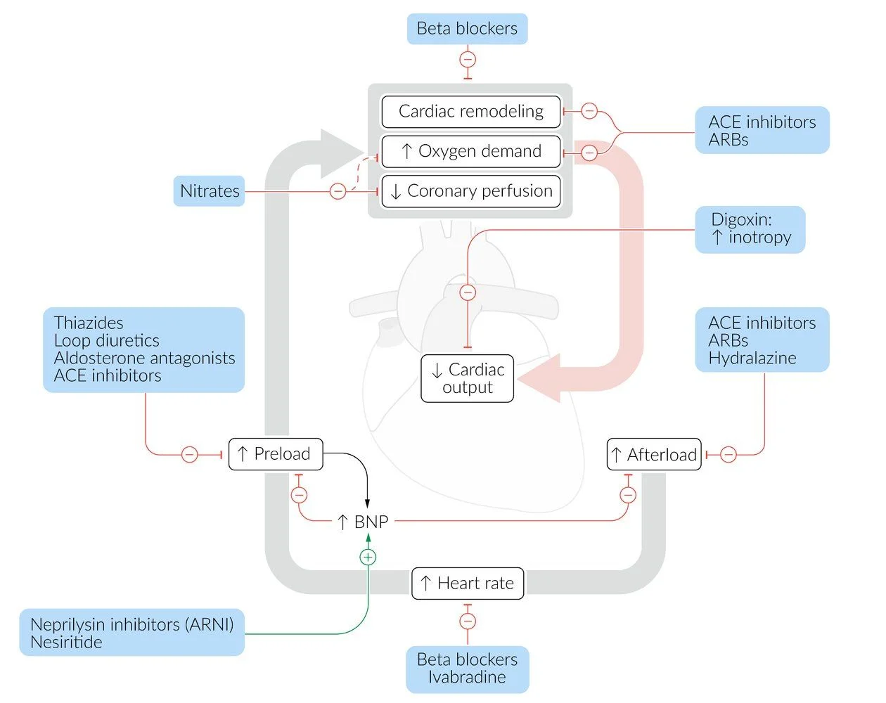
What this shows
Special case, asymptomatic EF ≤40% (NYHA I)
start an ACEi/ARB, then a β-blocker; this prevents progression to symptomatic HF.| Therapy | Mortality benefit | Indication |
|---|---|---|
| Beta blocker (metoprolol succinate, carvedilol, bisoprolol) | Yes | All HFrEF |
| ARNI (sacubitril-valsartan), or ACE inhibitor or ARB | Yes | All HFrEF; ARNI preferred |
| Aldosterone antagonist (spironolactone, eplerenone) | Yes | Persistent symptoms on initial therapy |
| SGLT2 inhibitor (dapagliflozin) | Yes | Persistent symptoms; all HFrEF with diabetes |
| Hydralazine plus isosorbide dinitrate | Yes | Intolerance of angiotensin-converting enzyme (ACE) inhibitor, angiotensin receptor blocker (ARB) or ARNI, or persistent symptoms |
| Loop diuretic | No (symptoms and hospitalization) | Volume overload |
| Digoxin | No (hospitalization only) | Persistent symptoms, especially with atrial fibrillation |
| Ivabradine | No | Sinus rhythm, pulse over 70 on maximal beta blocker |
| Implantable cardioverter-defibrillator (ICD) | Yes | NYHA II to III with ejection fraction under 35 percent, or aborted cardiac arrest |
| Cardiac resynchronization therapy (CRT) | Yes | Ejection fraction under 35 percent with QRS of 150 ms or more, or 120 ms or more with left bundle branch block |
| Cardiac transplant | Yes | Refractory HF despite maximal therapy |
HFpEF, drugs to avoid and prognosis
HFpEF, non-pharmacological measures
as for HFrEF; weight loss matters most here, because obesity and inactivity are stronger risk factors for HFpEF than for HFrEF.HFpEF, first line
an SGLT2 inhibitor for every patient, and a loop diuretic when volume overloaded.- Additional: an ARNI or ARB (candesartan) and an MRA (spironolactone).
- Control blood pressure to reduce afterload, and treat exacerbating conditions (CAD, obstructive sleep apnea, atrial fibrillation); exercise training and cardiac rehabilitation.
Drugs that worsen HF (avoid)
they can precipitate acute failure.- Non-dihydropyridine calcium-channel blockers.
- Non-steroidal anti-inflammatory drugs (NSAIDs) (they cause weight gain and fluid retention).
- Thiazolidinediones (pioglitazone), and tricyclic antidepressants.
Poor prognostic signs (asked on NBME)
any sign of worsening cardiac function.- Congestion (edema, S3, crackles).
- A persistently high BNP or N-terminal pro-BNP (NT-proBNP).
- Hyponatremia, systolic blood pressure under 120 mm Hg, an ICD in place.
- Unintentional weight loss, and comorbid diabetes, anemia or obesity.
Acute decompensation is its own emergency
Acute Decompensated Heart Failure.Which four drug classes improve survival in HFrEF, and which two commonly used drugs relieve symptoms or admissions without improving survival?
The four pillars: an ARNI (or ACE inhibitor/ARB), an evidence-based beta blocker (carvedilol, metoprolol succinate, bisoprolol), an MRA (spironolactone or eplerenone) and an SGLT2 inhibitor. Loop diuretics (symptoms and hospitalization) and digoxin (hospitalization only) do not improve survival.
How it's tested
Acute dyspnea with orthopnea, JVD, S3 and diffuse crackles, BP 150/90: acute decompensated heart failure — IV furosemide while hunting for the precipitant.
Severe hypertension, sudden respiratory distress, pink frothy sputum: hypertensive flash pulmonary edema — NIPPV and IV nitroglycerin now, then diuresis; do not wait for the film.
Cool extremities, BP 78/50, oliguria, confusion and pulmonary congestion: cardiogenic shock — inotrope and vasopressor, not a bigger diuretic dose.
An obese patient with convincing congestion and a BNP of 80: obesity lowers BNP; treat the picture.
A patient admitted for the third time this year: medication and diet non-adherence is the most common precipitant — ask before adding drugs.
Stable HFrEF, EF 30%, on furosemide alone: add the four pillars — the diuretic controls volume but changes no outcomes.
EF 25%, NYHA III, sinus rhythm, LBBB with QRS 165 ms on maximal drugs: CRT-D.
Newly diagnosed HFrEF admitted with pulmonary edema, BP 95/60, cool extremities: stabilize and diurese first; the beta blocker starts once compensated.
Black patient with NYHA III symptoms on all four pillars: add hydralazine + isosorbide dinitrate.
Serum sodium 127 in advanced HFrEF with edema: dilutional hyponatremia — diurese and restrict fluid; salt tablets are wrong.
Persistent EF 30% ten days after an anterior STEMI: optimize GDMT and re-echo at 40 days before considering an ICD.
A 74-year-old hypertensive woman with exertional dyspnea, edema, an S4, concentric LVH and EF 60%: HFpEF — loop diuretic for congestion plus an SGLT2 inhibitor; control blood pressure.
Which drug improves outcomes in HFpEF: an SGLT2 inhibitor; the beta blocker is for her AF, not her EF.
Obese patient with orthopnea, echo evidence of elevated filling pressure and a low-normal BNP: still HFpEF — obesity suppresses BNP.
Preserved EF with a pericardial knock, prior chest radiation and a sharp y descent: constrictive pericarditis — pericardiectomy, not generic HF drugs.
Preserved EF, thick walls, low ECG voltage, proteinuria and carpal tunnel: cardiac amyloidosis — biopsy or PYP scan; avoid digoxin and calcium-channel blockers.
Go deeper
Guidelines: 2022 AHA/ACC/HFSA Heart Failure Guideline · 2024 ACC HFrEF Decision Pathway · 2026 ACC HFpEF Decision Pathway
Related Step 2 pages: Heart Failure, Cor Pulmonale, High Output Heart Failure, Cardiorenal Syndrome, Dilated Cardiomyopathy, Approach to Shock
High-yield images4
Flashcards for this page
Card 1 of 5 · try-out only, nothing is saved
You just read one page of 236
The rest of Step 2 CK, written exactly like this.
Rapid Review is the reading layer of a full Step 1 platform: a schedule fitted to your exam date, flashcards for every page, and a mentor a message away.
Every Step 1 system
Written the same way: what to know, how it’s tested, where to go deeper.
A plan built to your exam date
Tell us when you sit, and the schedule fits the whole library and your question bank to it.
Flashcards for every page
Ready-made spaced-repetition decks linked to each topic, so nothing you read gets forgotten.
