
Rapid Review·Cardiovascular
Select any text to highlight it or add a note.
Foundations
T2High yieldJugular Venous Pressure
Focus on
Read the JVP in two steps: estimate right atrial pressure from the column height, then match the abnormal wave to its mechanical event — cannon a waves mean AV dissociation, giant v waves mean tricuspid regurgitation, and the y descent separates constriction from tamponade.
Key takeaways
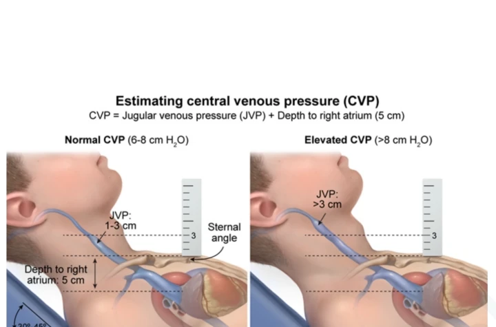
Estimating central venous pressure
What this shows
Find the internal jugular pulsation
recline the patient 30 to 45°.- It is biphasic and non-palpable.
- It falls with inspiration and rises with abdominal pressure.
- A carotid pulse does none of these things.
Measure, then add 5
the vertical height of the top of the column above the sternal angle, plus ~5 cm for the distance to the right atrium (RA).- Normal is ≤ 8 to 9 cm H2O; a column more than 3 cm above the sternal angle is elevated.
- See the JVP pressure estimation figure.
An elevated jugular venous pressure (JVP) means raised right-sided filling pressure
right or biventricular heart failure, tricuspid disease, pericardial constriction or tamponade, pulmonary hypertension, volume overload, superior vena cava (SVC) obstruction.The normal waveform
What this shows
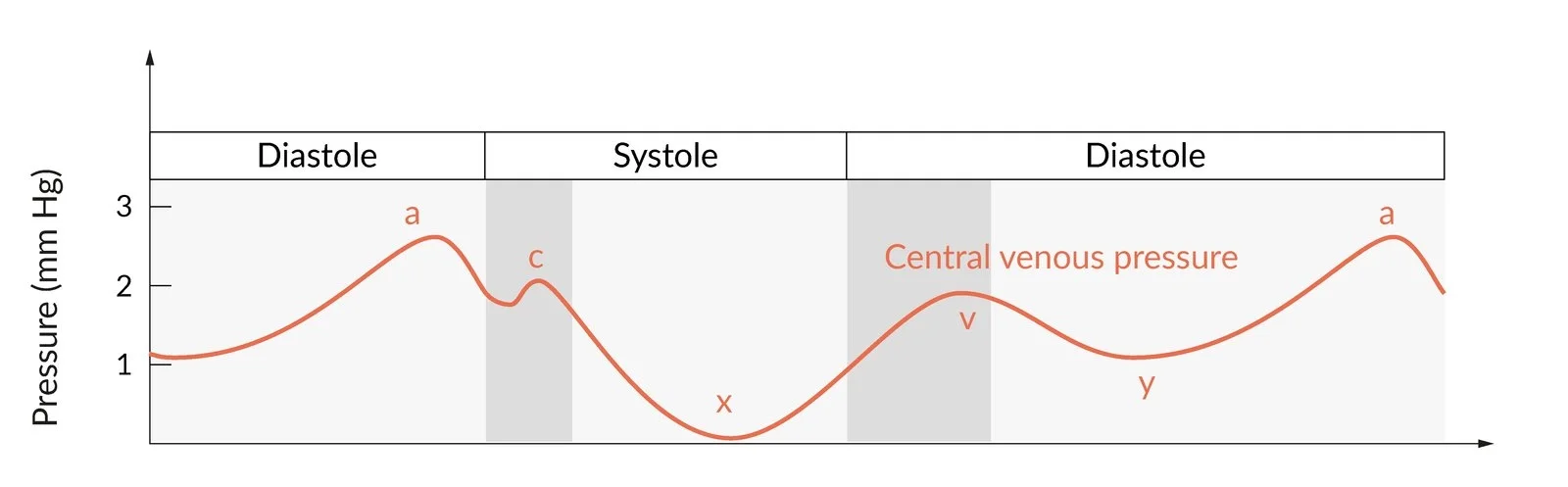
Five parts, each a mechanical event
see the normal waveform.a wave
right atrial contraction (late diastole).c wave
tricuspid valve bulging into the RA at the start of systole.x descent
atrial relaxation and downward movement of the tricuspid annulus during systole.v wave
venous filling of the atrium against a closed tricuspid valve (late systole).y descent
the tricuspid valve opens and the atrium empties (early diastole).Abnormal waveforms
What this shows
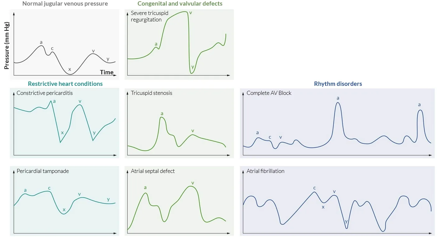
| Pattern | Mechanism | Diagnosis |
|---|---|---|
| Absent a waves | No organized atrial contraction | Atrial Fibrillation |
| Cannon a waves (intermittent, giant) | Atrium contracts against a closed tricuspid valve, atrioventricular (AV) dissociation | Atrioventricular Block, Ventricular Tachycardia, frequent premature ventricular contractions (PVCs) |
| Large regular a waves | High resistance to right ventricular filling | Tricuspid stenosis, pulmonary hypertension, pulmonic stenosis |
| Giant v waves, absent x descent | Systolic regurgitation into the RA | Tricuspid Regurgitation |
| v wave ≥ a wave | Left-to-right shunt volume | Atrial Septal Defect |
| Prominent x and y descents | Rapid early filling then abrupt halt | Constrictive Pericarditis |
| Prominent x, blunted or absent y | Filling impaired throughout diastole | Cardiac Tamponade |
Constriction and tamponade share a high JVP
in both the pressure outside the atria is raised.- The y descent separates them: sharp in constriction, blunted or absent in tamponade.
Cannon a waves can be felt
patients sometimes report a pulsing sensation in the neck.Respiration and the Kussmaul sign
Normal JVP falls with inspiration
a JVP that rises or fails to fall on inspiration is Kussmaul sign.- Causes: constrictive pericarditis, restrictive cardiomyopathy, right ventricular infarction, severe right heart failure, massive pulmonary embolism (PE).
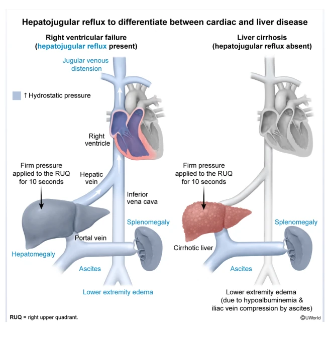
Abdominojugular (hepatojugular) reflux
What this shows
The test
firm pressure over the abdomen for 10 to 15 s while watching the neck.- A sustained rise ≥ 3 cm means the right heart cannot accommodate the extra venous return: heart failure or high filling pressures.
- A brief rise that fades is normal.
It separates cardiac from hepatic edema
the bedside maneuver for edema and ascites.- Positive in heart failure.
- Negative in uncomplicated cirrhosis.
A patient with a heart rate of 38 has intermittent giant pulsations in the neck. Name the wave, the mechanism and the likely rhythm.
Cannon a waves. The atrium contracts against a closed tricuspid valve because atria and ventricles beat independently (AV dissociation). With a rate of 38 the likely rhythm is complete heart block.
How it's tested
A patient with a heart rate of 38 and intermittent giant pulsations in the neck: cannon a waves from complete heart block — atria contracting against a closed tricuspid valve.
Holosystolic murmur louder on inspiration with prominent neck-vein pulsations and a pulsatile liver: tricuspid regurgitation; the v wave is the regurgitant jet.
Elevated JVP that rises on inspiration in a patient with prior chest radiation, ascites and pericardial calcification: constrictive pericarditis with Kussmaul sign — expect a sharp y descent.
Leg edema and ascites; the resting JVP looks borderline: press on the abdomen — a sustained ≥ 3 cm rise points to the heart, not the liver.
Go deeper
Related Step 2 pages: Heart Sounds & Murmurs, Constrictive Pericarditis, Cardiac Tamponade, Tricuspid Regurgitation, Cor Pulmonale, Cardiac Tamponade
High-yield images6
Flashcards for this page
Card 1 of 4 · try-out only, nothing is saved
You just read one page of 236
The rest of Step 2 CK, written exactly like this.
Rapid Review is the reading layer of a full Step 1 platform: a schedule fitted to your exam date, flashcards for every page, and a mentor a message away.
Every Step 1 system
Written the same way: what to know, how it’s tested, where to go deeper.
A plan built to your exam date
Tell us when you sit, and the schedule fits the whole library and your question bank to it.
Flashcards for every page
Ready-made spaced-repetition decks linked to each topic, so nothing you read gets forgotten.
