
Rapid Review·Cardiovascular
Select any text to highlight it or add a note.
Foundations
T2High yieldHeart Sounds & Murmurs
Videos
1 to play here
Play 1 here
Focus on
Turn a described sound into a diagnosis: the two gallops, the four S2 splits, each murmur by timing and location, and the bedside maneuvers — standing/Valsalva makes only HOCM and MVP louder, handgrip makes regurgitant murmurs louder and AS/HOCM softer.
Key takeaways
Extra heart sounds (gallops)
Third heart sound (S3)
just after the second heart sound (S2), rhythm "ken-TUC-ky".- Blood rushing into a dilated, volume-overloaded ventricle in early diastole.
- Pathologic in heart failure with reduced ejection fraction (HFrEF), high-output states (thyrotoxicosis, anemia), and mitral or aortic regurgitation (MR/AR).
- Can be normal in children, athletes, pregnancy and age under 40.
Fourth heart sound (S4)
just before the first heart sound (S1), rhythm "TEN-nes-see".- The atrium contracting into a stiff ventricle.
- Seen in concentric left ventricular hypertrophy (LVH) from hypertension or aortic stenosis (AS), heart failure with preserved ejection fraction (HFpEF), restrictive cardiomyopathy (CM), acute myocardial infarction (MI).
- It needs atrial contraction, so it disappears in atrial fibrillation.
| S3 | S4 | |
|---|---|---|
| Timing | Early diastole, after S2 | Late diastole, before S1 |
| Mechanism | Rapid filling of a dilated ventricle | Atrial kick into a non-compliant ventricle |
| Associations | HFrEF, MR/AR, high-output states | LVH, HFpEF, restrictive CM, acute MI |
| Can be normal? | Yes, young, athletes, pregnancy | Rarely (elderly) |
Splitting of S2
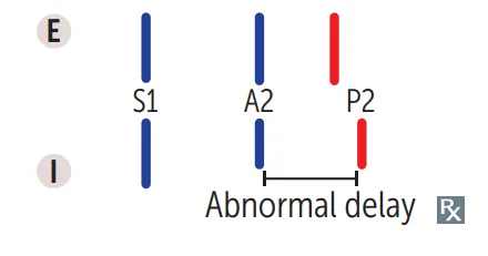
Four patterns
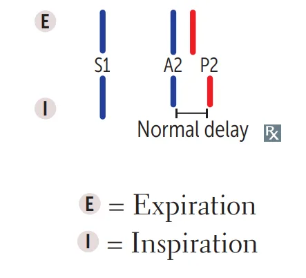
the Review S2 splitting patterns shows all four.Physiologic
inspiration lowers intrathoracic pressure, so more venous return and a longer right ventricular (RV) ejection delay P2.- Inspiration also raises pulmonary capacity (the pulmonary circulation holds more blood), which delays pulmonic closure further.
- The split widens on inspiration and closes on expiration.
Wide
anything that delays RV emptying: pulmonic stenosis, right bundle branch block (RBBB), pulmonary hypertension.- P2 comes markedly late, especially on inspiration; it still varies with breathing.
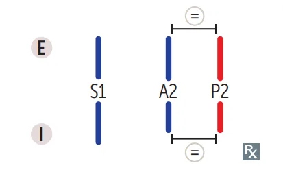
Fixed
Atrial Septal Defect (ASD).- The continuous left-to-right shunt keeps RV volume high in both phases, so the split is wide and never changes.
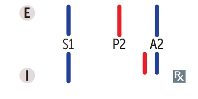
Paradoxical
aortic closure is delayed so P2 comes before A2: severe aortic stenosis, left bundle branch block (LBBB), hypertrophic obstructive cardiomyopathy (HOCM).- On inspiration P2 moves later, toward A2, so the split disappears.
- The split is heard on expiration, the opposite of normal.
What this shows
What this shows
Systolic murmurs
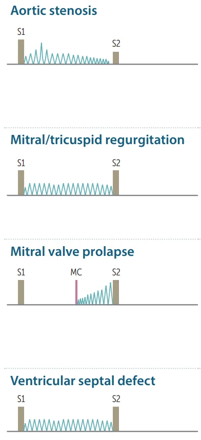
What this shows
Ejection vs holosystolic
ejection murmurs (crescendo-decrescendo) leave S1 audible; holosystolic murmurs bury S1.- Mitral regurgitation is blowing; a VSD is harsh: both are holosystolic.
- Where each area sits is on the auscultation areas.
| Murmur | Sound and site | Clues |
|---|---|---|
| Aortic Stenosis | Crescendo-decrescendo, right upper sternal border (RUSB), loudest at the base, radiates to carotids; soft/single S2, ± ejection click | Pulsus parvus et tardus; syncope, angina, dyspnea |
| Mitral Regurgitation | Holosystolic, high-pitched blowing, apex, radiating to the axilla | Post-MI, mitral valve prolapse (MVP), dilated left ventricle, rheumatic |
| Tricuspid Regurgitation | Holosystolic at the left lower sternal border (LLSB), louder on inspiration | Large v waves, pulsatile liver, RV dilation, endocarditis |
| Mitral Valve Prolapse | Mid-systolic click then late systolic murmur, apex, loudest just before S2; the click comes after the carotid pulse | Click moves earlier with standing/Valsalva |
| Ventricular Septal Defect (VSD) | Holosystolic, harsh, LLSB (tricuspid area) with thrill | Smaller defect = louder |
| Hypertrophic Cardiomyopathy | Ejection murmur at LLSB, no carotid radiation | Louder with less preload (standing, Valsalva) |
| Pulmonic flow / Pulmonary Stenosis | Ejection murmur at the left upper sternal border (LUSB) ± click, widens S2 | Congenital; Noonan, carcinoid |
Diastolic and continuous murmurs (always pathologic)
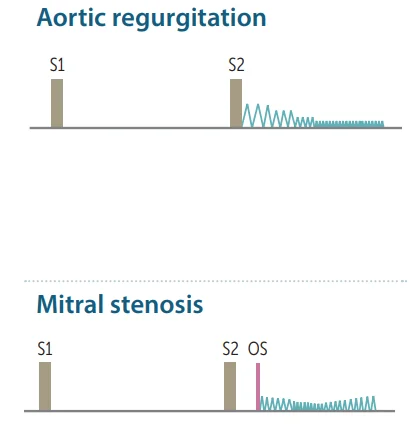
What this shows
Aortic regurgitation
early diastolic, decrescendo, blowing (Aortic Regurgitation).- Heard at the left sternal border (third left space, valve disease) or the right base (second right space, root dilation).
- Wide pulse pressure, bounding pulses, head bobbing.
- Louder with handgrip and leaning forward on expiration.
Mitral stenosis
opening snap (OS) then a low-pitched mid-diastolic rumble at the apex (Mitral Stenosis).- Heard with the bell, patient in the left lateral decubitus position.
- Almost always rheumatic.
- A shorter S2 to OS gap means more severe stenosis.
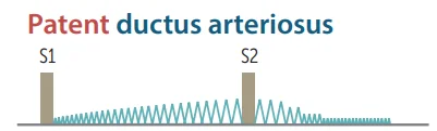
What this shows
Patent ductus arteriosus
continuous machinery murmur, left infraclavicular, loudest at S2; bounding pulses (Patent Ductus Arteriosus).Not a murmur
a triphasic scratchy rub that tracks the heartbeat and is louder leaning forward is a pericardial friction rub (Acute Pericarditis).Bedside maneuvers (the exam favorite)
One rule covers most of the table
preload and afterload move most murmurs one way and HOCM and MVP the other.| Maneuver | Physiology | Louder | Softer |
|---|---|---|---|
| Standing, Valsalva strain | Less preload, smaller left ventricle | HOCM, MVP (click earlier) | Almost everything else |
| Squatting, passive leg raise | More preload (and afterload with squatting) | Most murmurs, AS, MR, AR, VSD | HOCM, MVP (click later) |
| Sustained handgrip | More afterload | MR, AR, VSD | AS, HOCM |
| Inspiration | More right-sided venous return | Right-sided murmurs (tricuspid regurgitation, TR; pulmonic stenosis, PS) | Left-sided murmurs |
| Amyl nitrite / vasodilator | Less afterload | AS, HOCM, MVP | MR, AR, VSD |
Pressure tracings and when to get an echo
Pressure tracings
compare the Compare valve-lesion pressure curves.- Stenosis creates a pressure gradient across the valve.
- Regurgitation volume-loads the chamber upstream.
When a murmur needs an echocardiogram
any of these.- Any diastolic or continuous murmur.
- Any holosystolic or grade ≥3 murmur.
- A murmur with symptoms, abnormal S2, click, or abnormal pulses.
When it needs nothing
soft mid-systolic murmurs in an asymptomatic patient with a normal exam need nothing further.- The pediatric version of that rule is on Approach to Pediatric Murmurs.
- Innocent murmurs get softer on standing; the venous hum is the exception (louder standing, abolished by neck rotation or jugular pressure).
A systolic murmur gets louder when the patient stands up and softer when the patient squats. Which two lesions do this, and why?
HOCM and mitral valve prolapse. Standing lowers preload and the left ventricle gets smaller, which makes both louder (and moves the prolapse click earlier). Squatting raises preload and afterload, so both get softer while most other murmurs get louder.
How it's tested
A systolic murmur that gets louder when the patient stands or strains: only HOCM and MVP do that; everything else softens. Squatting reverses it.
Fixed splitting of S2 with an ejection murmur at the left upper sternal border: atrial septal defect — the murmur is pulmonic flow, not the shunt.
An opening snap and a diastolic rumble in a young immigrant woman with dyspnea in pregnancy: rheumatic mitral stenosis; the closer the snap to S2, the tighter the valve.
A holosystolic murmur at the left lower sternal border that gets louder with inspiration, with a pulsatile liver: tricuspid regurgitation.
Handgrip makes the murmur louder: a regurgitant left-sided lesion (MR, AR, VSD); it makes aortic stenosis and HOCM softer.
Go deeper
Guidelines: 2020 ACC/AHA Valvular Heart Disease Guideline
Related Step 2 pages: Jugular Venous Pressure, Aortic Stenosis, Mitral Regurgitation, Mitral Stenosis, Aortic Regurgitation, Hypertrophic Cardiomyopathy, Approach to Pediatric Murmurs
High-yield images10
Flashcards for this page
Card 1 of 3 · try-out only, nothing is saved
You just read one page of 240
The rest of Step 2 CK, written exactly like this.
Rapid Review is the reading layer of a full Step 1 platform: a schedule fitted to your exam date, flashcards for every page, and a mentor a message away.
Every Step 1 system
Written the same way: what to know, how it’s tested, where to go deeper.
A plan built to your exam date
Tell us when you sit, and the schedule fits the whole library and your question bank to it.
Flashcards for every page
Ready-made spaced-repetition decks linked to each topic, so nothing you read gets forgotten.
