
Rapid Review·Cardiovascular
Select any text to highlight it or add a note.
Shock
T2High yieldObstructive Shock
Focus on
Hypotension with distended neck veins and clear lungs — the heart cannot fill or empty because something mechanical is in the way. Three emergencies, three bedside fixes: tension pneumothorax (decompress on clinical grounds), tamponade (drain), massive PE (lyse). Fluids are a bridge; relieving the obstruction is the treatment.
Key takeaways
What it is and what causes it
Obstructive shock
a physical block to venous return, ventricular filling or outflow.- Output falls, vasoconstriction raises resistance, and systemic venous pressure rises (jugular venous distension (JVD)) while the left heart is under-filled.
Before the left heart
pulmonary embolism (PE) and tension pneumothorax give a high central venous pressure with a low or normal wedge pressure.Tamponade
equalizes diastolic pressures across all chambers, so it is the one obstructive shock with a high wedge pressure.The big three
tension pneumothorax (trauma, mechanical ventilation, central line, ruptured bleb), cardiac tamponade (malignancy, uremia, dissection, trauma, post-infarction rupture), massive PE.Other causes
abdominal compartment syndrome, auto positive end-expiratory pressure (PEEP) in ventilated asthma or chronic obstructive pulmonary disease (COPD), atrial myxoma, constrictive pericarditis, air embolism.Clinical features
Shared
hypotension, tachycardia, JVD, cool extremities, dyspnea and clear lungs (no pulmonary edema).Separating the three
by the bedside clues below.| Cause | Bedside clues | Immediate test | Definitive emergency treatment |
|---|---|---|---|
| Tension pneumothorax | Sudden dyspnea, unilateral absent breath sounds and hyperresonance, hypotension, JVD; tracheal deviation is late | Clinical diagnosis, no radiograph first; ultrasound shows absent lung sliding | Needle or finger decompression (5th intercostal space anterior axillary line, or 2nd space mid-clavicular), then chest tube |
| Cardiac Tamponade | Beck triad (hypotension, JVD, muffled sounds), pulsus paradoxus, narrow pulse pressure, electrical alternans | Bedside echo: effusion with right-sided diastolic collapse, plethoric inferior vena cava | Pericardiocentesis; surgery for trauma, dissection, rupture |
| Massive PE | Sudden dyspnea, syncope, pleuritic pain, hypoxemia, JVD, right-ventricular heave, loud P2, leg swelling | Bedside echo: dilated hypokinetic right ventricle, McConnell sign; CT angiography only if stable enough to travel | Systemic thrombolysis (alteplase 100 mg over 2 hours); catheter or surgical embolectomy if contraindicated or failed; anticoagulation |
Diagnosis
The point-of-care ultrasound sequence
four views.- Absent lung sliding means pneumothorax.
- Pericardial fluid with chamber collapse means tamponade.
- A dilated right ventricle with a small hyperdynamic left ventricle means PE.
- The inferior vena cava is plethoric in all three.
Electrocardiogram
sinus tachycardia, S1Q3T3 or new right bundle branch block (PE); low voltage and alternans (tamponade).No computed tomography (CT) for the unstable
decompress, drain or lyse on bedside evidence.Pulmonary artery catheter
high right-sided pressures with a low wedge in PE and pneumothorax; equalized diastolic pressures in tamponade.Management
Relieve the obstruction
(table above); everything else is a bridge.Fluids, cautiously
a 250 to 500 mL bolus can raise right-sided preload in tamponade or PE.- Large volumes over-distend the right ventricle, push the septum into the left ventricle and worsen output.
Norepinephrine
to maintain coronary perfusion while preparing definitive therapy.Avoid intubation and positive-pressure ventilation if possible
in tamponade and massive PE they cut venous return and can precipitate arrest; if unavoidable, have the definitive intervention ready.Arrest from obstruction
tamponade, tension pneumothorax and PE are three of the reversible T's; decompress, drain or give thrombolytics during resuscitation (Cardiac Arrest & ACLS).Versus cardiogenic shock
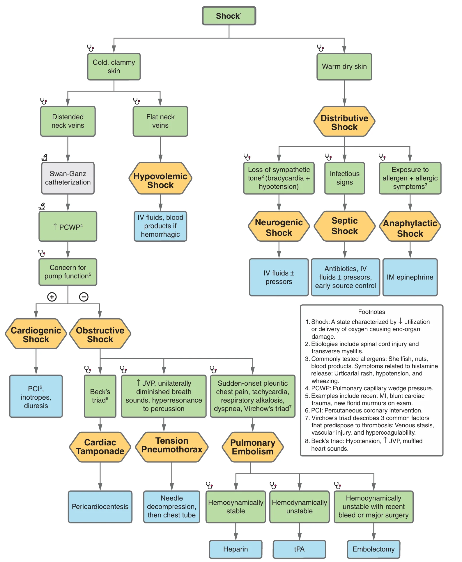
What this shows
Both have JVD and low output
but pulmonary edema with a high wedge means left-ventricular pump failure, while clear lungs with acute right-ventricular findings mean PE, tamponade or pneumothorax.The overlap
the right-ventricular infarct (inferior infarction, JVD, clear lungs) gets fluids and reperfusion (Cardiogenic Shock).A ventilated trauma patient suddenly becomes hypotensive with distended neck veins, absent breath sounds on the right and a hyperresonant right chest. What is the diagnosis, and should a chest radiograph come first?
Tension pneumothorax, a clinical diagnosis: no radiograph first. Do immediate needle or finger decompression (5th intercostal space anterior axillary line, or 2nd space mid-clavicular), then place a chest tube.
How it's tested
A ventilated trauma patient suddenly drops his pressure; the right chest is silent and hyperresonant, neck veins bulge: tension pneumothorax — decompress now; a chest film is a fatal delay.
Cancer patient with syncope, BP 80/50, clear lungs and a dilated RV on bedside echo: massive PE with obstructive shock — systemic thrombolysis, not routine anticoagulation and a CT.
Uremic patient with hypotension, JVD, muffled sounds and a 20 mm Hg pulsus paradoxus: tamponade — echo-guided pericardiocentesis.
Hemodynamics: CVP 20, PCWP 6, cardiac index 1.8, SVR high: obstruction before the left heart — PE or tension pneumothorax, not LV failure.
Intubating a tamponade patient for "respiratory distress" leads to arrest: positive pressure removed the last of the venous return — drain first, or at the same time.
Go deeper
Guidelines: 2026 ACC/AHA Acute Pulmonary Embolism Guideline · 2025 AHA Adult Advanced Life Support Guidance
Related Step 2 pages: Approach to Shock, Cardiac Tamponade, Cardiogenic Shock, Cardiac Arrest & ACLS, Cor Pulmonale
High-yield images1
Flashcards for this page
Card 1 of 2 · try-out only, nothing is saved
You just read one page of 236
The rest of Step 2 CK, written exactly like this.
Rapid Review is the reading layer of a full Step 1 platform: a schedule fitted to your exam date, flashcards for every page, and a mentor a message away.
Every Step 1 system
Written the same way: what to know, how it’s tested, where to go deeper.
A plan built to your exam date
Tell us when you sit, and the schedule fits the whole library and your question bank to it.
Flashcards for every page
Ready-made spaced-repetition decks linked to each topic, so nothing you read gets forgotten.
