
Rapid Review·Cardiovascular
Select any text to highlight it or add a note.
Arrhythmias: Ventricular & Arrest
T2High yieldCardiac Arrest & ACLS
Focus on
No pulse after 10 seconds means CPR now and a rhythm check in two-minute cycles. VF/pulseless VT gets shocks, epinephrine every 3–5 minutes and amiodarone after the third shock; PEA/asystole gets epinephrine at once and a hunt through the H's and T's. For a tachycardia with a pulse the only questions are stable or not, narrow or wide, regular or not.
Key takeaways
What it is and what causes it
Sudden cardiac arrest
the sudden loss of effective cardiac output: global hypoperfusion, loss of consciousness, no pulse; unreversed, it becomes sudden cardiac death.Most common cause in adults
a non-perfusing ventricular arrhythmia (ventricular tachycardia (VT) or ventricular fibrillation (VF)).Less common
pulseless electrical activity (PEA) and asystole, which reflect severe systemic illness (hypoxia, pulmonary embolism).- PEA is an organized rhythm with no pump action (electromechanical dissociation).
Causes of VT and VF
four groups.- Ischemia: an acute myocardial infarction is the primary trigger (VF is the most common cause of myocardial infarction (MI)-related death), chronic scar, heart failure with an ejection fraction under 30 to 35 percent, severe aortic stenosis.
- Structural: hypertrophic cardiomyopathy, an anomalous coronary artery, myocarditis, amyloid or sarcoid infiltration.
- Channelopathies: long QT (torsades), Brugada, and Wolff-Parkinson-White (pre-excited atrial fibrillation degenerating into VF).
- Commotio cordis: a chest blow during repolarization triggers VF.
Causes of PEA and asystole (the Hs and Ts)
ten causes.- Hypovolemia, hypoxemia, hydrogen ions (acidosis), hyper- or hypokalemia, hypothermia.
- Tension pneumothorax, tamponade, toxins (opioids, beta blockers), thrombosis (pulmonary or coronary), trauma.
Diagnosis and the arrest rhythms
Diagnosis
unresponsive with no pulse after 10 seconds of carotid or radial palpation; a monitor then shows the rhythm.Automated external defibrillator
does not display the rhythm; it shocks only if it detects VT or VF.| Rhythm | Appearance | Shockable |
|---|---|---|
| Ventricular fibrillation | Coarse or fine irregular waves of varying size and shape, no isoelectric line | Yes |
| Pulseless VT | Rapid, regular broad complexes (monomorphic, polymorphic or torsades) | Yes |
| Pulseless electrical activity | Any organized rhythm (often slow, wide, distorted) without a pulse; diagnosed after excluding VF and VT | No |
| Asystole | A gently undulating flat line, no electrical activity | No |
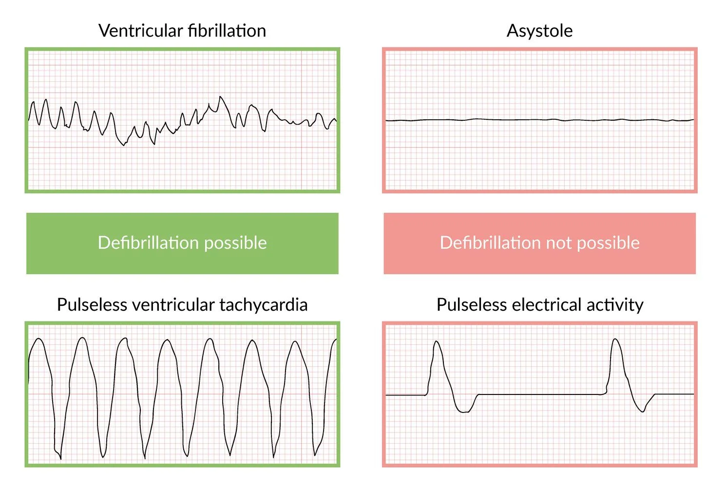
What this shows
Advanced cardiac life support
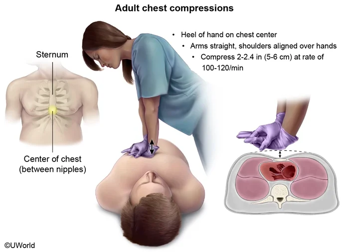
What this shows
All rhythms first (advanced cardiac life support, ACLS)
start high-quality chest compressions at once, ventilate at 10 to 12 breaths per minute, attach the defibrillator, and identify the rhythm, as in the Adult cardiac arrest pathway.Cycle every 2 minutes
cardiopulmonary resuscitation (CPR), then a rhythm and pulse check; shock if shockable; adjust as the rhythm changes.Shockable (VT or VF)
in order.- Immediate defibrillation: then 2 minutes of CPR.
- Epinephrine: 1 mg intravenously every 3 to 5 minutes.
- Amiodarone: 300 mg after the third shock.
Non-shockable (PEA or asystole)
uninterrupted CPR, no defibrillation, epinephrine 1 mg at once and then every 3 to 5 minutes, and a search for the reversible cause.| Cause | Fix |
|---|---|
| Hypovolemia | Saline bolus |
| Hypoxemia | Oxygenation |
| Hyperkalemia | Calcium chloride |
| Tension pneumothorax | Needle decompression |
| Tamponade | Pericardiocentesis |
| Massive pulmonary embolism | Thrombolysis |
PEA after an infarct
shock with chest pain and PEA 5 days after an infarct, with clear lungs, is a free-wall rupture with hemorrhagic tamponade.Tachyarrhythmia with a pulse
Instability means synchronized cardioversion
the shock is timed to the QRS so it does not land on repolarization and cause VF, as in the Tachyarrhythmia with a pulse pathway.- Instability from the rate itself is uncommon below 150 per minute.
Stable wide-complex tachycardia
the drug depends on the rhythm.- Amiodarone, procainamide, lidocaine or sotalol for monomorphic VT.
- Magnesium for torsades.
- Procainamide for pre-excited atrial fibrillation.
Prognosis
The most important factor
the time to effective resuscitation (early bystander CPR and early defibrillation).Worse outcome
three groups.- An initial rhythm of PEA or asystole; survival rises from asystole (the lowest chance of return of circulation) to PEA to VT or VF.
- CPR over 5 minutes, persistent coma, intubation or vasopressors.
- Older age and comorbidity (sepsis, stroke, class III or IV heart failure), and pneumonia or renal failure after CPR.
Better outcome
young healthy patients with a VT or VF arrest and early defibrillation.Survivors
get an implantable cardioverter-defibrillator.A man collapses; he has no pulse, and the monitor shows chaotic irregular waves with no isoelectric line. After the first shock and 2 minutes of CPR the rhythm is unchanged. What comes next, and when is amiodarone given?
Ventricular fibrillation, a shockable rhythm: shock again, continue CPR, and give epinephrine 1 mg every 3 to 5 minutes. Amiodarone 300 mg is given after the third shock. The biggest survival factor is the time to effective resuscitation.
How it's tested
A man collapses in the ward, no pulse, monitor shows VF: CPR, defibrillate, CPR 2 minutes, shock, epinephrine, shock, amiodarone.
Organized sinus rhythm at 90 on the monitor but no pulse in a trauma patient with a distended abdomen: PEA from hypovolemia — CPR, epinephrine, blood and fluids; no shock.
Pulseless arrest in a dialysis patient who missed two sessions: hyperkalemia — IV calcium plus insulin-glucose during CPR.
Post-operative day 3, sudden collapse, dilated RV on bedside echo: massive PE — thrombolysis during resuscitation.
Comatose after ROSC from VF with ST elevation in V2–V4: emergency coronary angiography plus targeted temperature management.
What single factor most improves survival after out-of-hospital arrest: early bystander CPR and defibrillation.
Go deeper
Guidelines: 2025 AHA Adult Advanced Life Support Guidance
Related Step 2 pages: Ventricular Tachycardia, Torsades de Pointes, Approach to Tachyarrhythmias, Cardiac Tamponade, Obstructive Shock, Cardiac Implantable Devices
High-yield images4
Flashcards for this page
Card 1 of 5 · try-out only, nothing is saved
You just read one page of 236
The rest of Step 2 CK, written exactly like this.
Rapid Review is the reading layer of a full Step 1 platform: a schedule fitted to your exam date, flashcards for every page, and a mentor a message away.
Every Step 1 system
Written the same way: what to know, how it’s tested, where to go deeper.
A plan built to your exam date
Tell us when you sit, and the schedule fits the whole library and your question bank to it.
Flashcards for every page
Ready-made spaced-repetition decks linked to each topic, so nothing you read gets forgotten.
