
Rapid Review·Cardiovascular
Select any text to highlight it or add a note.
Congenital Heart Disease
T1Must knowTetralogy of Fallot
Focus on
The most common cyanotic heart defect: pulmonary infundibular stenosis (the driver), RV hypertrophy, VSD and an overriding aorta. Cyanosis depends on how tight the outflow is; tet spells are treated with the knee-chest position, oxygen, morphine, fluids and a beta blocker; the boot-shaped heart and single S2 are the giveaways; surgical repair at 3–6 months.
Key takeaways
What it is
What this shows
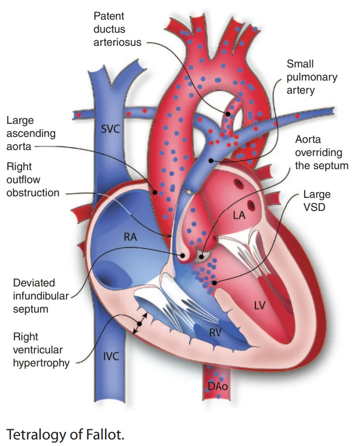
Tetralogy of Fallot
the most common cyanotic congenital heart disease, four defects from one malalignment (Cardiac Embryology Essentials).- Right ventricular outflow obstruction: pulmonary infundibular stenosis, the main anomaly.
- Right ventricular hypertrophy.
- A ventricular septal defect.
- An overriding aorta.
The degree of outflow obstruction determines everything
mild obstruction gives a left-to-right shunt across the defect and little cyanosis; severe obstruction gives a right-to-left shunt with deep cyanosis.Associations
typically sporadic; DiGeorge (22q11) and Down syndromes, and fetal alcohol exposure.Clinical features
Cyanosis within weeks of birth
mild to severe, with dyspnea on feeds and poor growth.Squatting
older children squat after exertion (it raises systemic resistance and pushes blood into the lungs).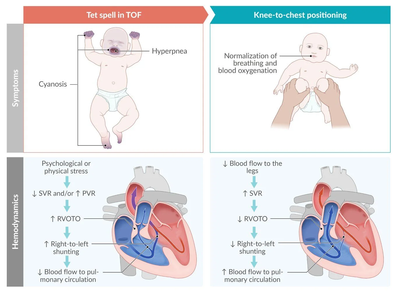
What this shows
Tet spells
intermittent hypercyanotic, hypoxic episodes, peak incidence 2 to 4 months, triggered by crying, feeding or defecation; squatting relieves them.The murmur
a harsh systolic ejection murmur at the left upper sternal border with a single S2 (sub-pulmonic stenosis; the large defect is silent), a right-ventricular heave and thrill.Diagnosis
Fetal echocardiography
diagnoses most cases; transthoracic echocardiography is confirmatory.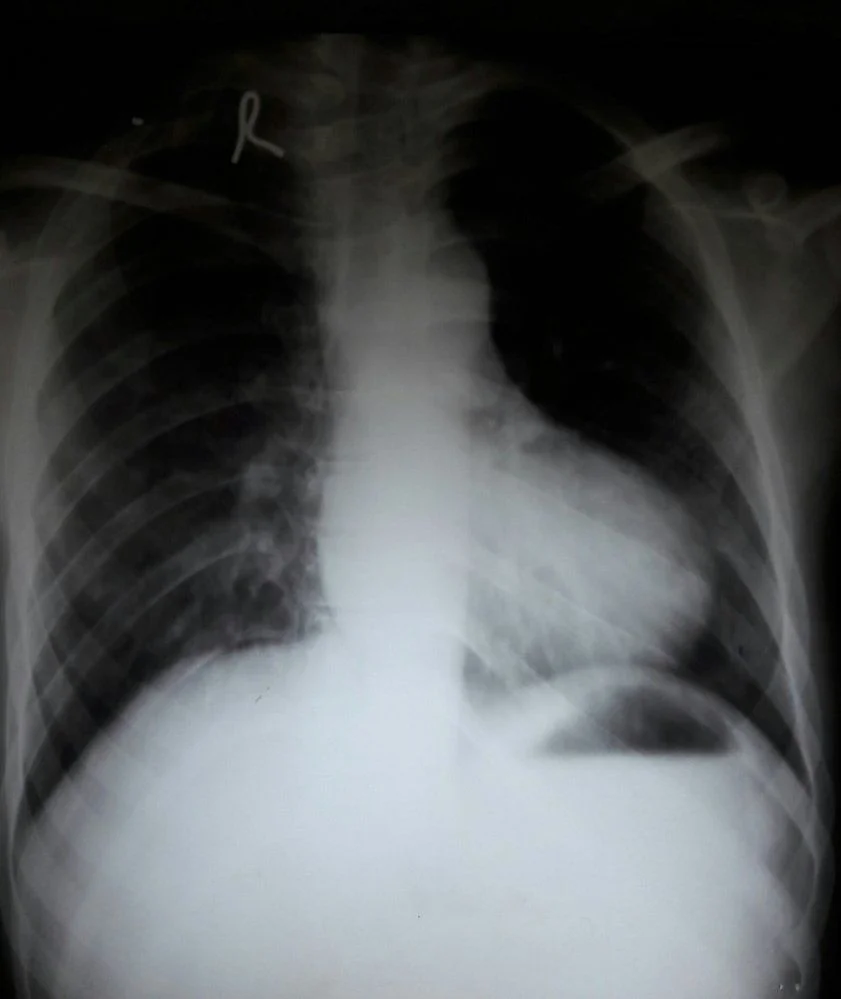
What this shows
Chest film
a boot-shaped heart with normal or decreased pulmonary vascular markings (the opposite of the shunt lesions).Electrocardiogram
right axis deviation and right ventricular hypertrophy.Hyperoxia test
fails (Approach to Neonatal Cyanosis).| Tetralogy of Fallot | Transposition of the Great Arteries | Truncus Arteriosus | Tricuspid Atresia | |
|---|---|---|---|---|
| Cyanosis onset | Weeks to months (spells at 2 to 4 months); at birth if severe | First hours | First weeks, with heart failure | First days |
| Murmur | Harsh ejection murmur at the left upper sternal border, single S2 | Usually none; single loud S2 | Harsh systolic, single S2, bounding pulses | Holosystolic, single S2 |
| Chest film | Boot-shaped heart, oligemic lungs | Egg on a string, plethoric lungs | Plethoric lungs, right arch | Oligemic lungs |
| Electrocardiogram | Right axis | Right ventricular hypertrophy | Biventricular hypertrophy | Left axis deviation |
| Association | DiGeorge, Down, fetal alcohol | Maternal diabetes | DiGeorge, maternal diabetes | Maternal diabetes |
Management
A tet spell, in order
step up until it breaks.- Knee-chest position and oxygen: the position raises systemic resistance and venous return; oxygen is a pulmonary vasodilator.
- Morphine: for sedation if unresponsive.
- Intravenous fluids and a beta blocker: if refractory, for volume, and to reduce sympathetic drive and infundibular spasm.
Other spell drugs
phenylephrine raises systemic resistance; avoid inotropes.Severe obstruction at birth
prostaglandin E1 infusion until urgent surgery (or a palliative shunt).Otherwise
elective surgical repair at 3 to 6 months (defect closure plus relief of the outflow obstruction); without surgery about half do not live past 3 years.Complications
spells with seizures or death, polycythemia, cerebral thrombosis and brain abscess (right-to-left shunt), endocarditis.- After repair: pulmonary regurgitation from the transannular patch with right-ventricular dilation (Pulmonary Regurgitation), arrhythmias including ventricular tachycardia and sudden death, right bundle branch block.
A 3-month-old turns deeply blue and breathes fast while crying; between episodes there is a harsh ejection murmur at the left upper sternal border with a single S2, which fades during the episode. What is happening, and what are the first two steps?
A tet spell in tetralogy of Fallot. First the knee-chest position and oxygen; then morphine if unresponsive, and intravenous fluids and a beta blocker if refractory. Definitive repair at 3 to 6 months.
How it's tested
A 3-month-old turns deeply blue and limp while crying; his usual murmur is barely audible; he improves when his mother holds him with his knees against his chest: tet spell — knee-chest position, oxygen, morphine, fluids, then a beta blocker; refer for early repair.
A cyanotic infant with a harsh ejection murmur at the left upper sternal border, a single S2 and a boot-shaped heart with dark lungs on the film: tetralogy of Fallot — echocardiogram; surgery at 3–6 months.
A 5-year-old immigrant who squats after running and has clubbed fingers: unrepaired tetralogy — squatting raises systemic resistance and reduces the right-to-left shunt.
Tetralogy patient with hypocalcemic seizures and no thymic shadow: 22q11.2 deletion (DiGeorge).
A 25-year-old with repaired tetralogy, worsening exercise tolerance, a diastolic murmur and a dilated RV on MRI: pulmonary regurgitation from the transannular patch — pulmonary valve replacement.
Which of the four defects determines the severity of cyanosis: the degree of right ventricular outflow obstruction.
Go deeper
Guidelines: 2018 AHA/ACC Adult Congenital Heart Disease Guideline
Related Step 2 pages: Approach to Neonatal Cyanosis, Approach to Congenital Heart Disease, Pulmonary Regurgitation, Pulmonary Stenosis, Transposition of the Great Arteries, Cardiac Embryology Essentials
High-yield images3
You just read one page of 236
The rest of Step 2 CK, written exactly like this.
Rapid Review is the reading layer of a full Step 1 platform: a schedule fitted to your exam date, flashcards for every page, and a mentor a message away.
Every Step 1 system
Written the same way: what to know, how it’s tested, where to go deeper.
A plan built to your exam date
Tell us when you sit, and the schedule fits the whole library and your question bank to it.
Flashcards for every page
Ready-made spaced-repetition decks linked to each topic, so nothing you read gets forgotten.
